 Movie
Movie Controller

[English] 日本語
 Yorodumi
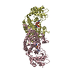
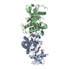
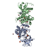
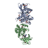
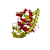
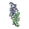
- PDB-7ev2: Crystal structure of Mycobacterium tuberculosis tryptophanyl-tRNA... -
+ Open data

- Basic information
| Entry | Database: PDB / ID: 7ev2 | ||||||
|---|---|---|---|---|---|---|---|
| Title | Crystal structure of Mycobacterium tuberculosis tryptophanyl-tRNA synthetase complexed with Y-11 and ATP | ||||||
 Components Components | Tryptophan--tRNA ligase | ||||||
 Keywords Keywords | LIGASE / TrpRS / aminoacylation / tRNA-binding / aminoacyl-tRNA synthetase / ATP-binding | ||||||
| Function / homology |  Function and homology information Function and homology informationtryptophan-tRNA ligase / tryptophanyl-tRNA aminoacylation / tryptophan-tRNA ligase activity / ATP binding / cytosol Similarity search - Function | ||||||
| Biological species |   Mycobacterium tuberculosis (bacteria) Mycobacterium tuberculosis (bacteria) | ||||||
| Method |  X-RAY DIFFRACTION / SYNCHROTRON / MOLECULAR REPLACEMENT / Resolution: 2.1 Å X-RAY DIFFRACTION / SYNCHROTRON / MOLECULAR REPLACEMENT / Resolution: 2.1 Å | ||||||
 Authors Authors | Xu, M. / Chen, S. | ||||||
 Citation Citation | Journal: Acs Chem.Biol. / Year: 2022 Title: Investigate Natural Product Indolmycin and the Synthetically Improved Analogue Toward Antimycobacterial Agents. Authors: Yang, Y. / Xu, Y. / Yue, Y. / Wang, H. / Cui, Y. / Pan, M. / Zhang, X. / Zhang, L. / Li, H. / Xu, M. / Tang, Y. / Chen, S. | ||||||
| History |
|
- Structure visualization
| Structure viewer | Molecule: MolmilJmol/JSmol |
|---|
- Downloads & links
-Download
| PDBx/mmCIF format | 7ev2.cif.gz | 85.7 KB | Display | PDBx/mmCIF format |
|---|---|---|---|---|
| PDB format | pdb7ev2.ent.gz | 60.1 KB | Display | PDB format |
| PDBx/mmJSON format | 7ev2.json.gz | Tree view | PDBx/mmJSON format | |
| Others |  Other downloads Other downloads |
-Validation report
| Arichive directory | https://data.pdbj.org/pub/pdb/validation_reports/ev/7ev2ftp://data.pdbj.org/pub/pdb/validation_reports/ev/7ev2 | HTTPS FTP |
|---|
-Related structure data
| Related structure data |  7el8SC 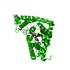 7eltC 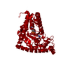 7ensC  7entC 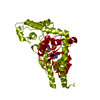 7ev3C S: Starting model for refinement C: citing same article ( |
|---|---|
| Similar structure data |


-Links
 PDBj
PDBj
- Assembly
| Deposited unit | 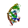
| ||||||||||||
|---|---|---|---|---|---|---|---|---|---|---|---|---|---|
| 1 | 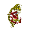
| ||||||||||||
| Unit cell |
| ||||||||||||
| Components on special symmetry positions |
|
-Components
| #1: Protein | Mass: 37380.992 Da / Num. of mol.: 1 Source method: isolated from a genetically manipulated source Source: (gene. exp.) Mycobacterium tuberculosis (bacteria)Gene: trpS, DSI38_02915, E5M05_20150, E5M52_19725, E5M78_19780, ERS007657_01660, ERS007661_00440, ERS007663_02313, ERS007665_01484, ERS007670_03000, ERS007679_02115, ERS007681_00969, ERS007688_01638, ...Gene: trpS, DSI38_02915, E5M05_20150, E5M52_19725, E5M78_19780, ERS007657_01660, ERS007661_00440, ERS007663_02313, ERS007665_01484, ERS007670_03000, ERS007679_02115, ERS007681_00969, ERS007688_01638, ERS007703_02455, ERS007722_03290, ERS007741_01346, ERS013471_03442, ERS023446_03614, ERS024276_00235, ERS027659_00823, ERS094182_03915, F6W99_01991, GCL30_20825, SAMEA2683035_03669 Production host: |
|---|---|
| #2: Chemical | ChemComp-ATP /   Mass: 507.181 Da / Num. of mol.: 1 / Source method: obtained synthetically / Formula: C10H16N5O13P3 / Feature type: SUBJECT OF INVESTIGATION / Comment: ATP, energy-carrying molecule*YM Mass: 507.181 Da / Num. of mol.: 1 / Source method: obtained synthetically / Formula: C10H16N5O13P3 / Feature type: SUBJECT OF INVESTIGATION / Comment: ATP, energy-carrying molecule*YM |
| #3: Chemical | ChemComp-MG /   Mass: 24.305 Da / Num. of mol.: 1 / Source method: obtained synthetically / Formula: Mg / Feature type: SUBJECT OF INVESTIGATION Mass: 24.305 Da / Num. of mol.: 1 / Source method: obtained synthetically / Formula: Mg / Feature type: SUBJECT OF INVESTIGATION |
| #4: Chemical | ChemComp-JE0 / (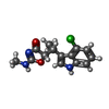  Mass: 291.733 Da / Num. of mol.: 1 / Source method: obtained synthetically / Formula: C14H14ClN3O2 / Feature type: SUBJECT OF INVESTIGATION Mass: 291.733 Da / Num. of mol.: 1 / Source method: obtained synthetically / Formula: C14H14ClN3O2 / Feature type: SUBJECT OF INVESTIGATION |
| #5: Water | ChemComp-HOH /  Mass: 18.015 Da / Num. of mol.: 202 / Source method: isolated from a natural source / Formula: H2O Mass: 18.015 Da / Num. of mol.: 202 / Source method: isolated from a natural source / Formula: H2O |
| Has ligand of interest | Y |
-Experimental details
-Experiment
| Experiment | Method: X-RAY DIFFRACTION / Number of used crystals: 1 |
|---|
- Sample preparation
| Crystal | Density Matthews: 2.45 Å3/Da / Density % sol: 49.87 % |
|---|---|
| Crystal grow | Temperature: 291 K / Method: vapor diffusion, sitting drop / pH: 5.7 / Details: 0.2M MgCl2, 35% PEG 400, 0.1M MES, pH 5.7 |
-Data collection
| Diffraction | Mean temperature: 100 K / Serial crystal experiment: N |
|---|---|
| Diffraction source | Source: SYNCHROTRON / Site: APS  / Beamline: 21-ID-D / Wavelength: 1.12713 Å / Beamline: 21-ID-D / Wavelength: 1.12713 Å |
| Detector | Type: DECTRIS EIGER X 9M / Detector: PIXEL / Date: Jul 26, 2019 |
| Radiation | Protocol: SINGLE WAVELENGTH / Monochromatic (M) / Laue (L): M / Scattering type: x-ray |
| Radiation wavelength | Wavelength: 1.12713 Å / Relative weight: 1 |
| Reflection | Resolution: 2.1→50 Å / Num. obs: 23421 / % possible obs: 99.9 % / Redundancy: 33.1 % / Biso Wilson estimate: 25.35 Å2 / CC1/2: 0.997 / Net I/σ(I): 41 |
| Reflection shell | Resolution: 2.1→2.14 Å / Redundancy: 36.2 % / Mean I/σ(I) obs: 6.8 / Num. unique obs: 1144 / CC1/2: 0.982 / % possible all: 100 |
- Processing
| Software |
| |||||||||||||||||||||||||||||||||||||||||||||||||||||||||||||||
|---|---|---|---|---|---|---|---|---|---|---|---|---|---|---|---|---|---|---|---|---|---|---|---|---|---|---|---|---|---|---|---|---|---|---|---|---|---|---|---|---|---|---|---|---|---|---|---|---|---|---|---|---|---|---|---|---|---|---|---|---|---|---|---|---|
| Refinement | Method to determine structure: MOLECULAR REPLACEMENT Starting model: 7EL8 Resolution: 2.1→35.64 Å / SU ML: 0.2348 / Cross valid method: FREE R-VALUE / σ(F): 1.34 / Phase error: 22.6844 Stereochemistry target values: GeoStd + Monomer Library + CDL v1.2
| |||||||||||||||||||||||||||||||||||||||||||||||||||||||||||||||
| Solvent computation | Shrinkage radii: 0.9 Å / VDW probe radii: 1.11 Å / Solvent model: FLAT BULK SOLVENT MODEL | |||||||||||||||||||||||||||||||||||||||||||||||||||||||||||||||
| Displacement parameters | Biso mean: 28 Å2 | |||||||||||||||||||||||||||||||||||||||||||||||||||||||||||||||
| Refinement step | Cycle: LAST / Resolution: 2.1→35.64 Å
| |||||||||||||||||||||||||||||||||||||||||||||||||||||||||||||||
| Refine LS restraints |
| |||||||||||||||||||||||||||||||||||||||||||||||||||||||||||||||
| LS refinement shell |
|
