 Movie
Movie Controller

[English] 日本語
 Yorodumi
- PDB-7eu9: Crystal structure of the selenomethionine(SeMet)-derived Cas12i1 ... -
+ Open data

- Basic information
| Entry | Database: PDB / ID: 7eu9 | ||||||
|---|---|---|---|---|---|---|---|
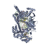
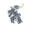
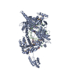
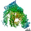
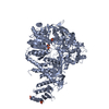
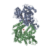
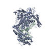
| Title | Crystal structure of the selenomethionine(SeMet)-derived Cas12i1 R-loop complex before target DNA cleavage | ||||||
 Components Components |
| ||||||
 Keywords Keywords | RNA BINDING PROTEIN / Cas12i / Cas12i1 / CRISPR | ||||||
| Function / homology | CITRIC ACID / DNA / DNA (> 10) / RNA / RNA (> 10) Function and homology information Function and homology information | ||||||
| Biological species |  Lachnospiraceae bacterium ND2006 (bacteria) Lachnospiraceae bacterium ND2006 (bacteria) | ||||||
| Method |  X-RAY DIFFRACTION / SYNCHROTRON / SAD / Resolution: 2.35 Å X-RAY DIFFRACTION / SYNCHROTRON / SAD / Resolution: 2.35 Å | ||||||
 Authors Authors | Zhang, B. / Luo, D.Y. / Li, Y. / OuYang, S.Y. | ||||||
| Funding support |  China, 1items China, 1items
| ||||||
 Citation Citation | Journal: Nat Commun / Year: 2021 Title: Mechanistic insights into the R-loop formation and cleavage in CRISPR-Cas12i1. Authors: Zhang, B. / Luo, D. / Li, Y. / Perculija, V. / Chen, J. / Lin, J. / Ye, Y. / Ouyang, S. | ||||||
| History |
|
- Structure visualization
| Structure viewer | Molecule: MolmilJmol/JSmol |
|---|
- Downloads & links
-Download
| PDBx/mmCIF format | 7eu9.cif.gz | 288.4 KB | Display | PDBx/mmCIF format |
|---|---|---|---|---|
| PDB format | pdb7eu9.ent.gz | 223.9 KB | Display | PDB format |
| PDBx/mmJSON format | 7eu9.json.gz | Tree view | PDBx/mmJSON format | |
| Others |  Other downloads Other downloads |
-Validation report
| Arichive directory | https://data.pdbj.org/pub/pdb/validation_reports/eu/7eu9ftp://data.pdbj.org/pub/pdb/validation_reports/eu/7eu9 | HTTPS FTP |
|---|
-Related structure data


-Links
 PDBj
PDBj


- Assembly
| Deposited unit | 
| ||||||||
|---|---|---|---|---|---|---|---|---|---|
| 1 |
| ||||||||
| Unit cell |
|
-Components
-DNA chain , 2 types, 2 molecules CD
| #3: DNA chain | Mass: 12326.935 Da / Num. of mol.: 1 / Source method: obtained synthetically Source: (synth.) Lachnospiraceae bacterium ND2006 (bacteria) |
|---|---|
| #4: DNA chain | Mass: 12241.883 Da / Num. of mol.: 1 / Source method: obtained synthetically Source: (synth.) Lachnospiraceae bacterium ND2006 (bacteria) |
-Protein / RNA chain , 2 types, 2 molecules AB
| #1: Protein | Mass: 128172.484 Da / Num. of mol.: 1 Source method: isolated from a genetically manipulated source Source: (gene. exp.) Lachnospiraceae bacterium ND2006 (bacteria)Production host: |
|---|---|
| #2: RNA chain | Mass: 13781.143 Da / Num. of mol.: 1 Source method: isolated from a genetically manipulated source Source: (gene. exp.) Lachnospiraceae bacterium ND2006 (bacteria)Production host: |
-Non-polymers , 2 types, 80 molecules 

| #5: Chemical | ChemComp-CIT /  Mass: 192.124 Da / Num. of mol.: 1 / Source method: obtained synthetically / Formula: C6H8O7 Mass: 192.124 Da / Num. of mol.: 1 / Source method: obtained synthetically / Formula: C6H8O7 |
|---|---|
| #6: Water | ChemComp-HOH / Mass: 18.015 Da / Num. of mol.: 79 / Source method: isolated from a natural source / Formula: H2O |
-Details
| Has ligand of interest | N |
|---|---|
| Has protein modification | Y |
-Experimental details
-Experiment
| Experiment | Method: X-RAY DIFFRACTION / Number of used crystals: 1 |
|---|
- Sample preparation
| Crystal | Density Matthews: 2.82 Å3/Da / Density % sol: 53.41 % |
|---|---|
| Crystal grow | Temperature: 289 K / Method: vapor diffusion, hanging drop Details: 0.1 M sodium citrate (pH 5.6), 17% (w/v) Polyethylene glycol 3350, 0.1 M sodium citrate |
-Data collection
| Diffraction | Mean temperature: 100 K / Serial crystal experiment: N |
|---|---|
| Diffraction source | Source: SYNCHROTRON / Site: SSRF / Beamline: BL17U1 / Wavelength: 0.9792 Å |
| Detector | Type: ADSC QUANTUM 315r / Detector: CCD / Date: Dec 30, 2019 |
| Radiation | Protocol: SINGLE WAVELENGTH / Monochromatic (M) / Laue (L): M / Scattering type: x-ray |
| Radiation wavelength | Wavelength: 0.9792 Å / Relative weight: 1 |
| Reflection | Resolution: 2.35→60 Å / Num. obs: 78375 / % possible obs: 100 % / Redundancy: 13.2 % / CC1/2: 0.997 / Rmerge(I) obs: 0.14 / Net I/σ(I): 9.4 |
| Reflection shell | Resolution: 2.35→2.48 Å / Rmerge(I) obs: 0.969 / Num. unique obs: 11343 / CC1/2: 0.83 |
- Processing
| Software |
| ||||||||||||||||||||||||||||||||||||||||||||||||||||||||||||
|---|---|---|---|---|---|---|---|---|---|---|---|---|---|---|---|---|---|---|---|---|---|---|---|---|---|---|---|---|---|---|---|---|---|---|---|---|---|---|---|---|---|---|---|---|---|---|---|---|---|---|---|---|---|---|---|---|---|---|---|---|---|
| Refinement | Method to determine structure: SAD / Resolution: 2.35→58.63 Å / Cor.coef. Fo:Fc: 0.93 / Cor.coef. Fo:Fc free: 0.906 / SU B: 8.487 / SU ML: 0.197 / Cross valid method: THROUGHOUT / σ(F): 0 / ESU R: 0.324 / ESU R Free: 0.246 / Stereochemistry target values: MAXIMUM LIKELIHOOD Details: HYDROGENS HAVE BEEN ADDED IN THE RIDING POSITIONS U VALUES : REFINED INDIVIDUALLY
| ||||||||||||||||||||||||||||||||||||||||||||||||||||||||||||
| Solvent computation | Ion probe radii: 0.8 Å / Shrinkage radii: 0.8 Å / VDW probe radii: 1.2 Å / Solvent model: MASK | ||||||||||||||||||||||||||||||||||||||||||||||||||||||||||||
| Displacement parameters | Biso max: 200 Å2 / Biso mean: 58.834 Å2 / Biso min: 27.49 Å2
| ||||||||||||||||||||||||||||||||||||||||||||||||||||||||||||
| Refinement step | Cycle: final / Resolution: 2.35→58.63 Å
| ||||||||||||||||||||||||||||||||||||||||||||||||||||||||||||
| Refine LS restraints |
| ||||||||||||||||||||||||||||||||||||||||||||||||||||||||||||
| LS refinement shell | Resolution: 2.35→2.411 Å / Rfactor Rfree error: 0 / Total num. of bins used: 20
|
