 Movie
Movie Controller

[English] 日本語
 Yorodumi
- PDB-7ete: Crystal structure of AbHpaI-Mg-(4R)-KDGal complex, Class II aldol... -
+ Open data

- Basic information
| Entry | Database: PDB / ID: 7ete | |||||||||
|---|---|---|---|---|---|---|---|---|---|---|
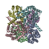
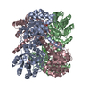
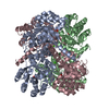
| Title | Crystal structure of AbHpaI-Mg-(4R)-KDGal complex, Class II aldolase, HpaI from Acinetobacter baumannii | |||||||||
 Components Components | 4-hydroxy-2-oxoheptanedioate aldolase | |||||||||
 Keywords Keywords | LYASE / TIM barrel / aldehyde lyase | |||||||||
| Function / homology |  Function and homology information Function and homology information: / 4-hydroxy-2-oxoheptanedioate aldolase / phenylacetate catabolic process / metal ion binding Similarity search - Function | |||||||||
| Biological species |  Acinetobacter baumannii (bacteria) Acinetobacter baumannii (bacteria) | |||||||||
| Method |  X-RAY DIFFRACTION / MOLECULAR REPLACEMENT / Resolution: 1.9 Å X-RAY DIFFRACTION / MOLECULAR REPLACEMENT / Resolution: 1.9 Å | |||||||||
| Model details | Crystal structure of AbHpaI-Zn-pyruvate complex, a class II aldolase, HpaI from Acinetobacter baumannii | |||||||||
 Authors Authors | Watthaisong, P. / Binlaeh, A. / Jaruwat, A. / Chaiyen, P. / Chitnumsub, P. / Maenpuen, S. | |||||||||
| Funding support |  Thailand, 2items Thailand, 2items
| |||||||||
 Citation Citation | Journal: J.Biol.Chem. / Year: 2021 Title: Catalytic and structural insights into a stereospecific and thermostable Class II aldolase HpaI from Acinetobacter baumannii. Authors: Watthaisong, P. / Binlaeh, A. / Jaruwat, A. / Lawan, N. / Tantipisit, J. / Jaroensuk, J. / Chuaboon, L. / Phonbuppha, J. / Tinikul, R. / Chaiyen, P. / Chitnumsub, P. / Maenpuen, S. | |||||||||
| History |
|
- Structure visualization
| Structure viewer | Molecule: MolmilJmol/JSmol |
|---|
- Downloads & links
-Download
| PDBx/mmCIF format | 7ete.cif.gz | 170.4 KB | Display | PDBx/mmCIF format |
|---|---|---|---|---|
| PDB format | pdb7ete.ent.gz | 132.9 KB | Display | PDB format |
| PDBx/mmJSON format | 7ete.json.gz | Tree view | PDBx/mmJSON format | |
| Others |  Other downloads Other downloads |
-Validation report
| Arichive directory | https://data.pdbj.org/pub/pdb/validation_reports/et/7eteftp://data.pdbj.org/pub/pdb/validation_reports/et/7ete | HTTPS FTP |
|---|
-Related structure data
| Related structure data |  7et8SC  7et9C 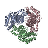 7etaC 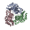 7etbC 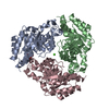 7etcC 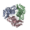 7etdC 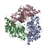 7etfC 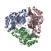 7etgC 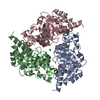 7ethC  7etiC S: Starting model for refinement C: citing same article ( |
|---|---|
| Similar structure data |


-Links
 PDBj
PDBj
- Assembly
| Deposited unit | 
| ||||||||
|---|---|---|---|---|---|---|---|---|---|
| 1 | 
| ||||||||
| Unit cell |
| ||||||||
| Components on special symmetry positions |
|
-Components
| #1: Protein | Mass: 28755.910 Da / Num. of mol.: 3 Source method: isolated from a genetically manipulated source Source: (gene. exp.) Acinetobacter baumannii (bacteria) / Gene: hpaI, FD887_17235 / Plasmid: pET11a / Production host: References: UniProt: A0A5R9HKL3, 4-hydroxy-2-oxoheptanedioate aldolase #2: Chemical |   Mass: 178.140 Da / Num. of mol.: 3 / Source method: obtained synthetically / Formula: C6H10O6 / Feature type: SUBJECT OF INVESTIGATION Mass: 178.140 Da / Num. of mol.: 3 / Source method: obtained synthetically / Formula: C6H10O6 / Feature type: SUBJECT OF INVESTIGATION#3: Chemical |   Mass: 24.305 Da / Num. of mol.: 3 / Source method: obtained synthetically / Formula: Mg Mass: 24.305 Da / Num. of mol.: 3 / Source method: obtained synthetically / Formula: Mg#4: Chemical | ChemComp-CA / |   Mass: 40.078 Da / Num. of mol.: 1 / Source method: obtained synthetically / Formula: Ca Mass: 40.078 Da / Num. of mol.: 1 / Source method: obtained synthetically / Formula: Ca#5: Water | ChemComp-HOH / |  Mass: 18.015 Da / Num. of mol.: 652 / Source method: isolated from a natural source / Formula: H2O Mass: 18.015 Da / Num. of mol.: 652 / Source method: isolated from a natural source / Formula: H2OHas ligand of interest | Y | |
|---|
-Experimental details
-Experiment
| Experiment | Method: X-RAY DIFFRACTION / Number of used crystals: 1 |
|---|
- Sample preparation
| Crystal | Density Matthews: 2.83 Å3/Da / Density % sol: 56.5 % |
|---|---|
| Crystal grow | Temperature: 277 K / Method: microbatch / pH: 4.6 / Details: CaCl2, MPD, Na acetate |
-Data collection
| Diffraction | Mean temperature: 100 K / Serial crystal experiment: N | ||||||||||||||||||||||||||||||
|---|---|---|---|---|---|---|---|---|---|---|---|---|---|---|---|---|---|---|---|---|---|---|---|---|---|---|---|---|---|---|---|
| Diffraction source | Source: ROTATING ANODE / Type: BRUKER TURBO X-RAY SOURCE / Wavelength: 1.54 Å | ||||||||||||||||||||||||||||||
| Detector | Type: BRUKER PHOTON 100 / Detector: CMOS / Date: Aug 7, 2020 | ||||||||||||||||||||||||||||||
| Radiation | Protocol: SINGLE WAVELENGTH / Monochromatic (M) / Laue (L): M / Scattering type: x-ray | ||||||||||||||||||||||||||||||
| Radiation wavelength | Wavelength: 1.54 Å / Relative weight: 1 | ||||||||||||||||||||||||||||||
| Reflection | Resolution: 1.8→24.41 Å / Num. obs: 89082 / % possible obs: 99.7 % / Redundancy: 6.7 % / CC1/2: 0.995 / Rmerge(I) obs: 0.121 / Rpim(I) all: 0.05 / Rrim(I) all: 0.131 / Net I/σ(I): 11 | ||||||||||||||||||||||||||||||
| Reflection shell | Diffraction-ID: 1
|
- Processing
| Software |
| ||||||||||||||||||||||||||||||||||||||||||||||||||||||||||||
|---|---|---|---|---|---|---|---|---|---|---|---|---|---|---|---|---|---|---|---|---|---|---|---|---|---|---|---|---|---|---|---|---|---|---|---|---|---|---|---|---|---|---|---|---|---|---|---|---|---|---|---|---|---|---|---|---|---|---|---|---|---|
| Refinement | Method to determine structure: MOLECULAR REPLACEMENT Starting model: 7ET8 Resolution: 1.9→24.4 Å / Cor.coef. Fo:Fc: 0.954 / Cor.coef. Fo:Fc free: 0.938 / SU B: 2.916 / SU ML: 0.083 / Cross valid method: THROUGHOUT / σ(F): 0 / ESU R: 0.123 / ESU R Free: 0.115 / Stereochemistry target values: MAXIMUM LIKELIHOOD Details: HYDROGENS HAVE BEEN ADDED IN THE RIDING POSITIONS U VALUES : REFINED INDIVIDUALLY
| ||||||||||||||||||||||||||||||||||||||||||||||||||||||||||||
| Solvent computation | Ion probe radii: 0.8 Å / Shrinkage radii: 0.8 Å / VDW probe radii: 1.2 Å / Solvent model: MASK | ||||||||||||||||||||||||||||||||||||||||||||||||||||||||||||
| Displacement parameters | Biso max: 67.97 Å2 / Biso mean: 15.856 Å2 / Biso min: 4.95 Å2
| ||||||||||||||||||||||||||||||||||||||||||||||||||||||||||||
| Refinement step | Cycle: final / Resolution: 1.9→24.4 Å
| ||||||||||||||||||||||||||||||||||||||||||||||||||||||||||||
| Refine LS restraints |
| ||||||||||||||||||||||||||||||||||||||||||||||||||||||||||||
| LS refinement shell | Resolution: 1.9→1.949 Å / Rfactor Rfree error: 0 / Total num. of bins used: 20
|
