 Movie
Movie Controller

[English] 日本語
 Yorodumi
- PDB-7eo5: Crystal structure of pyrano 1,3, oxazine derivative in complex wi... -
+ Open data

- Basic information
| Entry | Database: PDB / ID: 7eo5 | ||||||
|---|---|---|---|---|---|---|---|
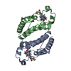
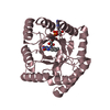
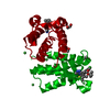
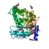
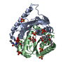
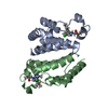
| Title | Crystal structure of pyrano 1,3, oxazine derivative in complex with the second bromodomain of BRD2 | ||||||
 Components Components | Bromodomain-containing protein 2 | ||||||
 Keywords Keywords | TRANSCRIPTION / BET family / BRD2 / bromodomain / b-mercaptoethanol / CME | ||||||
| Function / homology |  Function and homology information Function and homology informationacetylation-dependent protein binding / histone H3K14ac reader activity / chromatin looping / histone H4K5ac reader activity / histone H4K12ac reader activity / RUNX3 regulates p14-ARF / positive regulation of T-helper 17 cell lineage commitment / protein localization to chromatin / neural tube closure / nucleosome assembly ...acetylation-dependent protein binding / histone H3K14ac reader activity / chromatin looping / histone H4K5ac reader activity / histone H4K12ac reader activity / RUNX3 regulates p14-ARF / positive regulation of T-helper 17 cell lineage commitment / protein localization to chromatin / neural tube closure / nucleosome assembly / spermatogenesis / histone binding / nuclear speck / chromatin remodeling / protein serine/threonine kinase activity / chromatin binding / regulation of transcription by RNA polymerase II / chromatin / nucleoplasm / nucleus / cytoplasm Similarity search - Function | ||||||
| Biological species |  Homo sapiens (human) Homo sapiens (human) | ||||||
| Method |  X-RAY DIFFRACTION / MOLECULAR REPLACEMENT / Resolution: 2 Å X-RAY DIFFRACTION / MOLECULAR REPLACEMENT / Resolution: 2 Å | ||||||
 Authors Authors | Padmanabhan, B. / Arole, A. / Deshmukh, P. / Ashok, S. | ||||||
 Citation Citation | Journal: Acta Crystallogr.,Sect.F / Year: 2022 Title: Structural investigation of a pyrano-1,3-oxazine derivative and the phenanthridinone core moiety against BRD2 bromodomains. Authors: Arole, A.H. / Deshmukh, P. / Sridhar, A. / Padmanabhan, B. | ||||||
| History |
|
- Structure visualization
| Structure viewer | Molecule: MolmilJmol/JSmol |
|---|
- Downloads & links
-Download
| PDBx/mmCIF format | 7eo5.cif.gz | 51 KB | Display | PDBx/mmCIF format |
|---|---|---|---|---|
| PDB format | pdb7eo5.ent.gz | 27.9 KB | Display | PDB format |
| PDBx/mmJSON format | 7eo5.json.gz | Tree view | PDBx/mmJSON format | |
| Others |  Other downloads Other downloads |
-Validation report
| Arichive directory | https://data.pdbj.org/pub/pdb/validation_reports/eo/7eo5ftp://data.pdbj.org/pub/pdb/validation_reports/eo/7eo5 | HTTPS FTP |
|---|
-Related structure data
| Related structure data | 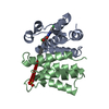 7envC 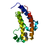 7enzC  5xheS S: Starting model for refinement C: citing same article ( |
|---|---|
| Similar structure data |

-Links
 PDBj
PDBj
- Assembly
| Deposited unit | 
| ||||||||||||
|---|---|---|---|---|---|---|---|---|---|---|---|---|---|
| 1 | 
| ||||||||||||
| Unit cell |
| ||||||||||||
| Components on special symmetry positions |
|
-Components
| #1: Protein | Mass: 13406.319 Da / Num. of mol.: 1 / Fragment: UNP RESIDUES 348-455 Source method: isolated from a genetically manipulated source Source: (gene. exp.) Homo sapiens (human) / Gene: BRD2, KIAA9001, RING3 / Plasmid: pET28a / Production host:  |
|---|---|
| #2: Chemical | ChemComp-PGE /   Mass: 150.173 Da / Num. of mol.: 1 / Source method: obtained synthetically / Formula: C6H14O4 Mass: 150.173 Da / Num. of mol.: 1 / Source method: obtained synthetically / Formula: C6H14O4 |
| #3: Chemical | ChemComp-BUX /   Mass: 325.104 Da / Num. of mol.: 1 / Source method: obtained synthetically / Formula: C13H6Cl2N2O4 / Feature type: SUBJECT OF INVESTIGATION Mass: 325.104 Da / Num. of mol.: 1 / Source method: obtained synthetically / Formula: C13H6Cl2N2O4 / Feature type: SUBJECT OF INVESTIGATION |
| #4: Water | ChemComp-HOH /  Mass: 18.015 Da / Num. of mol.: 132 / Source method: isolated from a natural source / Formula: H2O Mass: 18.015 Da / Num. of mol.: 132 / Source method: isolated from a natural source / Formula: H2O |
| Has ligand of interest | Y |
-Experimental details
-Experiment
| Experiment | Method: X-RAY DIFFRACTION / Number of used crystals: 1 |
|---|
- Sample preparation
| Crystal | Density Matthews: 2.2 Å3/Da / Density % sol: 44.12 % |
|---|---|
| Crystal grow | Temperature: 293 K / Method: vapor diffusion, hanging drop / pH: 7.5 / Details: 50 mM Tris pH 7.5, 50 mM NaCl, 30% PEG MME 2000 |
-Data collection
| Diffraction | Mean temperature: 100 K / Serial crystal experiment: N |
|---|---|
| Diffraction source | Source: ROTATING ANODE / Type: RIGAKU MICROMAX-007 HF / Wavelength: 1.54056 Å |
| Detector | Type: RIGAKU HyPix-6000HE / Detector: PIXEL / Date: Oct 15, 2020 |
| Radiation | Protocol: SINGLE WAVELENGTH / Monochromatic (M) / Laue (L): M / Scattering type: x-ray |
| Radiation wavelength | Wavelength: 1.54056 Å / Relative weight: 1 |
| Reflection | Resolution: 1.72→50 Å / Num. obs: 13233 / % possible obs: 98.5 % / Redundancy: 3.3 % / Biso Wilson estimate: 17.57 Å2 / CC1/2: 0.98 / Rmerge(I) obs: 0.078 / Net I/σ(I): 23.02 |
| Reflection shell | Resolution: 1.72→1.75 Å / Rmerge(I) obs: 0.26 / Num. unique obs: 610 / CC1/2: 0.86 |
- Processing
| Software |
| ||||||||||||||||||||||||
|---|---|---|---|---|---|---|---|---|---|---|---|---|---|---|---|---|---|---|---|---|---|---|---|---|---|
| Refinement | Method to determine structure: MOLECULAR REPLACEMENT Starting model: 5XHE Resolution: 2→29.07 Å / SU ML: 0.1701 / Cross valid method: FREE R-VALUE / σ(F): 1.34 / Phase error: 19.8388 Stereochemistry target values: GeoStd + Monomer Library + CDL v1.2 Details: Refinement was performed up to 2.0 A resolution.
| ||||||||||||||||||||||||
| Solvent computation | Shrinkage radii: 0.9 Å / VDW probe radii: 1.11 Å / Solvent model: FLAT BULK SOLVENT MODEL | ||||||||||||||||||||||||
| Displacement parameters | Biso mean: 19.47 Å2 | ||||||||||||||||||||||||
| Refinement step | Cycle: LAST / Resolution: 2→29.07 Å
| ||||||||||||||||||||||||
| Refine LS restraints |
| ||||||||||||||||||||||||
| LS refinement shell |
|
