 Movie
Movie Controller


+ Open data

- Basic information
| Entry | Database: PDB / ID: 7els | ||||||
|---|---|---|---|---|---|---|---|
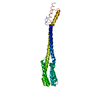
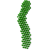
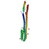
| Title | Crystal structure of xanthine riboswitch with 8-azaxanthine | ||||||
 Components Components | NMT1_8-azaxanthine (46-MER) | ||||||
 Keywords Keywords | RNA / riboswitch / 8-azaxanthine | ||||||
| Function / homology | 8-AZAXANTHINE / GUANOSINE-5'-TRIPHOSPHATE / RNA / RNA (> 10) Function and homology information Function and homology information | ||||||
| Biological species |  Ideonella sp. B508-1 (bacteria) Ideonella sp. B508-1 (bacteria) | ||||||
| Method |  X-RAY DIFFRACTION / SYNCHROTRON / MOLECULAR REPLACEMENT / Resolution: 3 Å X-RAY DIFFRACTION / SYNCHROTRON / MOLECULAR REPLACEMENT / Resolution: 3 Å | ||||||
 Authors Authors | Xu, X.C. / Ren, A.M. | ||||||
| Funding support |  China, 1items China, 1items
| ||||||
 Citation Citation | Journal: Nucleic Acids Res. / Year: 2021 Title: Insights into xanthine riboswitch structure and metal ion-mediated ligand recognition. Authors: Xu, X. / Egger, M. / Chen, H. / Bartosik, K. / Micura, R. / Ren, A. | ||||||
| History |
|
- Structure visualization
| Structure viewer | Molecule: MolmilJmol/JSmol |
|---|
- Downloads & links
-Download
| PDBx/mmCIF format | 7els.cif.gz | 67 KB | Display | PDBx/mmCIF format |
|---|---|---|---|---|
| PDB format | pdb7els.ent.gz | 47.2 KB | Display | PDB format |
| PDBx/mmJSON format | 7els.json.gz | Tree view | PDBx/mmJSON format | |
| Others |  Other downloads Other downloads |
-Validation report
| Arichive directory | https://data.pdbj.org/pub/pdb/validation_reports/el/7elsftp://data.pdbj.org/pub/pdb/validation_reports/el/7els | HTTPS FTP |
|---|
-Related structure data
| Related structure data |  7elpSC  7elqC 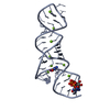 7elrC S: Starting model for refinement C: citing same article ( |
|---|---|
| Similar structure data |


-Links
 PDBj
PDBj


- Assembly
| Deposited unit | 
| ||||||||
|---|---|---|---|---|---|---|---|---|---|
| 1 | 
| ||||||||
| Unit cell |
|
-Components
| #1: RNA chain | Mass: 14558.750 Da / Num. of mol.: 2 Source method: isolated from a genetically manipulated source Source: (gene. exp.) Ideonella sp. B508-1 (bacteria)Production host: in vitro transcription vector pT7-TP(deltai) (others) #2: Chemical |   Mass: 523.180 Da / Num. of mol.: 2 / Source method: obtained synthetically / Formula: C10H16N5O14P3 / Comment: GTP, energy-carrying molecule*YM Mass: 523.180 Da / Num. of mol.: 2 / Source method: obtained synthetically / Formula: C10H16N5O14P3 / Comment: GTP, energy-carrying molecule*YM#3: Chemical | ChemComp-MG /   Mass: 24.305 Da / Num. of mol.: 13 / Source method: obtained synthetically / Formula: Mg / Feature type: SUBJECT OF INVESTIGATION Mass: 24.305 Da / Num. of mol.: 13 / Source method: obtained synthetically / Formula: Mg / Feature type: SUBJECT OF INVESTIGATION#4: Chemical |   Mass: 153.099 Da / Num. of mol.: 2 / Source method: obtained synthetically / Formula: C4H3N5O2 / Feature type: SUBJECT OF INVESTIGATION Mass: 153.099 Da / Num. of mol.: 2 / Source method: obtained synthetically / Formula: C4H3N5O2 / Feature type: SUBJECT OF INVESTIGATION#5: Water | ChemComp-HOH / |  Mass: 18.015 Da / Num. of mol.: 46 / Source method: isolated from a natural source / Formula: H2O Mass: 18.015 Da / Num. of mol.: 46 / Source method: isolated from a natural source / Formula: H2OHas ligand of interest | Y | |
|---|
-Experimental details
-Experiment
| Experiment | Method: X-RAY DIFFRACTION / Number of used crystals: 1 |
|---|
- Sample preparation
| Crystal | Density Matthews: 2.55 Å3/Da / Density % sol: 51.78 % |
|---|---|
| Crystal grow | Temperature: 289 K / Method: vapor diffusion, sitting drop Details: 0.1 M MES, pH 6.0, 0.2 M calcium acetate, 10% (v/v) isopropanol |
-Data collection
| Diffraction | Mean temperature: 100 K / Serial crystal experiment: N | |||||||||||||||||||||||||||||||||||||||||||||||||||||||||||||||||||||||||||||||||||||||||||||||||||
|---|---|---|---|---|---|---|---|---|---|---|---|---|---|---|---|---|---|---|---|---|---|---|---|---|---|---|---|---|---|---|---|---|---|---|---|---|---|---|---|---|---|---|---|---|---|---|---|---|---|---|---|---|---|---|---|---|---|---|---|---|---|---|---|---|---|---|---|---|---|---|---|---|---|---|---|---|---|---|---|---|---|---|---|---|---|---|---|---|---|---|---|---|---|---|---|---|---|---|---|---|
| Diffraction source | Source: SYNCHROTRON / Site: SSRF / Beamline: BL19U1 / Wavelength: 1.102 Å | |||||||||||||||||||||||||||||||||||||||||||||||||||||||||||||||||||||||||||||||||||||||||||||||||||
| Detector | Type: DECTRIS PILATUS 6M / Detector: PIXEL / Date: Nov 19, 2020 | |||||||||||||||||||||||||||||||||||||||||||||||||||||||||||||||||||||||||||||||||||||||||||||||||||
| Radiation | Protocol: SINGLE WAVELENGTH / Monochromatic (M) / Laue (L): M / Scattering type: x-ray | |||||||||||||||||||||||||||||||||||||||||||||||||||||||||||||||||||||||||||||||||||||||||||||||||||
| Radiation wavelength | Wavelength: 1.102 Å / Relative weight: 1 | |||||||||||||||||||||||||||||||||||||||||||||||||||||||||||||||||||||||||||||||||||||||||||||||||||
| Reflection | Resolution: 2.81→50 Å / Num. obs: 7789 / % possible obs: 98.6 % / Redundancy: 11.7 % / Rmerge(I) obs: 0.136 / Rpim(I) all: 0.043 / Rrim(I) all: 0.143 / Χ2: 1.318 / Net I/σ(I): 4 / Num. measured all: 91238 | |||||||||||||||||||||||||||||||||||||||||||||||||||||||||||||||||||||||||||||||||||||||||||||||||||
| Reflection shell | Diffraction-ID: 1
|
- Processing
| Software |
| ||||||||||||||||||||||||
|---|---|---|---|---|---|---|---|---|---|---|---|---|---|---|---|---|---|---|---|---|---|---|---|---|---|
| Refinement | Method to determine structure: MOLECULAR REPLACEMENT Starting model: 7ELP Resolution: 3→40.05 Å / SU ML: 0.35 / Cross valid method: THROUGHOUT / σ(F): 1.38 / Phase error: 36.07 / Stereochemistry target values: ML
| ||||||||||||||||||||||||
| Solvent computation | Shrinkage radii: 0.9 Å / VDW probe radii: 1.11 Å / Solvent model: FLAT BULK SOLVENT MODEL | ||||||||||||||||||||||||
| Displacement parameters | Biso max: 134.9 Å2 / Biso mean: 84.6421 Å2 / Biso min: 54.4 Å2 | ||||||||||||||||||||||||
| Refinement step | Cycle: final / Resolution: 3→40.05 Å
| ||||||||||||||||||||||||
| LS refinement shell | Refine-ID: X-RAY DIFFRACTION / Rfactor Rfree error: 0 / Total num. of bins used: 2 / % reflection obs: 99 %
|
