| 登録情報 | データベース: PDB / ID: 7ekw
|
|---|
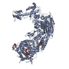
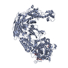
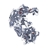
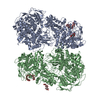
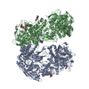
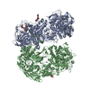
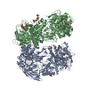
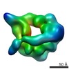
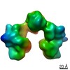
| タイトル | Crystal Structure of the Candida Glabrata Glycogen Debranching Enzyme (D535N) in complex with maltotetrose |
|---|
 要素 要素 | 4-alpha-glucanotransferase |
|---|
 キーワード キーワード | SUGAR BINDING PROTEIN / Glycogen Debranching Enzyme |
|---|
| 機能・相同性 |  機能・相同性情報 機能・相同性情報
amylo-alpha-1,6-glucosidase / amylo-alpha-1,6-glucosidase activity / 4-alpha-glucanotransferase / 4-alpha-glucanotransferase activity / glycogen biosynthetic process / glycogen catabolic process / cytoplasm類似検索 - 分子機能 Glycogen debranching enzyme, metazoa / Glycogen debranching enzyme / Eukaryotic glycogen debranching enzyme, N-terminal domain / Glycogen debranching enzyme, central domain / Glycogen debranching enzyme, C-terminal / Glycogen debranching enzyme, glucanotransferase domain / Amylo-alpha-1,6-glucosidase / N-terminal domain from the human glycogen debranching enzyme / Glycogen debranching enzyme, glucanotransferase domain / Central domain of human glycogen debranching enzyme ...Glycogen debranching enzyme, metazoa / Glycogen debranching enzyme / Eukaryotic glycogen debranching enzyme, N-terminal domain / Glycogen debranching enzyme, central domain / Glycogen debranching enzyme, C-terminal / Glycogen debranching enzyme, glucanotransferase domain / Amylo-alpha-1,6-glucosidase / N-terminal domain from the human glycogen debranching enzyme / Glycogen debranching enzyme, glucanotransferase domain / Central domain of human glycogen debranching enzyme / Six-hairpin glycosidase-like superfamily / Six-hairpin glycosidase superfamily / Glycoside hydrolase superfamily類似検索 - ドメイン・相同性 |
|---|
| 生物種 |  Candida glabrata CBS 138 (菌類) Candida glabrata CBS 138 (菌類) |
|---|
| 手法 |  X線回折 / シンクロトロン / 分子置換 / 解像度: 3.1 Å X線回折 / シンクロトロン / 分子置換 / 解像度: 3.1 Å |
|---|
 データ登録者 データ登録者 | Shen, M. / Xiang, S. |
|---|
| 資金援助 |  中国, 2件 中国, 2件 | 組織 | 認可番号 | 国 |
|---|
| National Natural Science Foundation of China (NSFC) | 31870769 | 中国 | | National Natural Science Foundation of China (NSFC) | 32071205 | 中国 |
|
|---|
 引用 引用 | ジャーナル: Acta Crystallogr.,Sect.F / 年: 2021
タイトル: Crystal structures of glycogen-debranching enzyme mutants in complex with oligosaccharides.
著者: Shen, M. / Gong, X. / Xiang, S. |
|---|
| 履歴 | | 登録 | 2021年4月7日 | 登録サイト: PDBJ / 処理サイト: PDBJ |
|---|
| 改定 1.0 | 2021年11月10日 | Provider: repository / タイプ: Initial release |
|---|
| 改定 1.1 | 2023年11月29日 | Group: Data collection / Refinement description
カテゴリ: chem_comp_atom / chem_comp_bond / pdbx_initial_refinement_model |
|---|
|
|---|
 ムービー
ムービー コントローラー


 データを開く
 基本情報
基本情報 構造の表示
構造の表示 ダウンロードとリンク
 その他のダウンロード
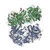


 PDBj
PDBj
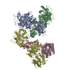
 集合体
集合体

 試料調製
試料調製 解析
解析