 Movie
Movie Controller

[English] 日本語
 Yorodumi
- PDB-7eji: Crystal structure of KRED F147L/L153Q/Y190P/L199A/M205F/M206F var... -
+ Open data

- Basic information
| Entry | Database: PDB / ID: 7eji | ||||||
|---|---|---|---|---|---|---|---|
| Title | Crystal structure of KRED F147L/L153Q/Y190P/L199A/M205F/M206F variant and methyl methacrylate complex | ||||||
 Components Components | 3-alpha-(Or 20-beta)-hydroxysteroid dehydrogenase | ||||||
 Keywords Keywords | OXIDOREDUCTASE / Ketoreductases / NADPH-dependent / Enantioselectivity | ||||||
| Function / homology |  Function and homology information Function and homology informationOxidoreductases; Acting on the CH-OH group of donors; With NAD+ or NADP+ as acceptor / oxidoreductase activity / nucleotide binding / metal ion binding Similarity search - Function | ||||||
| Biological species |  Lactobacillus kefiri (bacteria) Lactobacillus kefiri (bacteria) | ||||||
| Method |  X-RAY DIFFRACTION / SYNCHROTRON / MOLECULAR REPLACEMENT / Resolution: 1.56001602495 Å X-RAY DIFFRACTION / SYNCHROTRON / MOLECULAR REPLACEMENT / Resolution: 1.56001602495 Å | ||||||
 Authors Authors | Cui, J. / Huang, X. / Wang, B. / Zhao, H. / Zhou, J. | ||||||
 Citation Citation | Journal: Nat Catal / Year: 2022 Title: Photoinduced chemomimetic biocatalysis for enantioselective intermolecular radical conjugate addition Authors: Huang, X. / Feng, J. / Cui, J. / Jiang, G. / Harrison, W. / Zang, X. / Zhou, J. / Wang, B. / Zhao, H. | ||||||
| History |
|
- Structure visualization
| Structure viewer | Molecule: MolmilJmol/JSmol |
|---|
- Downloads & links
-Download
| PDBx/mmCIF format | 7eji.cif.gz | 239.4 KB | Display | PDBx/mmCIF format |
|---|---|---|---|---|
| PDB format | pdb7eji.ent.gz | 175.2 KB | Display | PDB format |
| PDBx/mmJSON format | 7eji.json.gz | Tree view | PDBx/mmJSON format | |
| Others |  Other downloads Other downloads |
-Validation report
| Summary document | 7eji_validation.pdf.gz | 2.2 MB | Display | wwPDB validaton report |
|---|---|---|---|---|
| Full document | 7eji_full_validation.pdf.gz | 2.2 MB | Display | |
| Data in XML | 7eji_validation.xml.gz | 45.8 KB | Display | |
| Data in CIF | 7eji_validation.cif.gz | 64.4 KB | Display | |
| Arichive directory | https://data.pdbj.org/pub/pdb/validation_reports/ej/7ejiftp://data.pdbj.org/pub/pdb/validation_reports/ej/7eji | HTTPS FTP |
-Related structure data
| Related structure data | 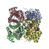 7ejhC 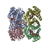 7ejjC 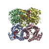 7vdoC 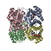 7ve7C 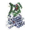 4rf2S S: Starting model for refinement C: citing same article ( |
|---|---|
| Similar structure data |
-Links
 PDBj
PDBj


- Assembly
| Deposited unit | 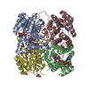
| ||||||||||||
|---|---|---|---|---|---|---|---|---|---|---|---|---|---|
| 1 |
| ||||||||||||
| Unit cell |
| ||||||||||||
| Components on special symmetry positions |
|
-Components
-Protein , 1 types, 4 molecules ABCD
| #1: Protein | Mass: 26773.164 Da / Num. of mol.: 4 / Mutation: F147L,L153Q,Y190P,L199A, M205F, M206F Source method: isolated from a genetically manipulated source Source: (gene. exp.) Lactobacillus kefiri (bacteria) / Gene: adhR, fabG3, DNL43_05835, LKE01_04370Production host: References: UniProt: Q6WVP7 |
|---|
-Non-polymers , 9 types, 663 molecules 


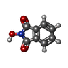
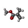


| #2: Chemical |  Mass: 24.305 Da / Num. of mol.: 2 / Source method: obtained synthetically / Formula: Mg Mass: 24.305 Da / Num. of mol.: 2 / Source method: obtained synthetically / Formula: Mg#3: Chemical |  Mass: 209.240 Da / Num. of mol.: 2 / Source method: obtained synthetically / Formula: C8H19NO5 / Comment: pH buffer*YM Mass: 209.240 Da / Num. of mol.: 2 / Source method: obtained synthetically / Formula: C8H19NO5 / Comment: pH buffer*YM#4: Chemical |  Mass: 92.094 Da / Num. of mol.: 3 / Source method: obtained synthetically / Formula: C3H8O3 Mass: 92.094 Da / Num. of mol.: 3 / Source method: obtained synthetically / Formula: C3H8O3#5: Chemical | ChemComp-NAP /  Mass: 743.405 Da / Num. of mol.: 4 / Source method: obtained synthetically / Formula: C21H28N7O17P3 / Feature type: SUBJECT OF INVESTIGATION Mass: 743.405 Da / Num. of mol.: 4 / Source method: obtained synthetically / Formula: C21H28N7O17P3 / Feature type: SUBJECT OF INVESTIGATION#6: Chemical |  Mass: 163.130 Da / Num. of mol.: 2 / Source method: obtained synthetically / Formula: C8H5NO3 Mass: 163.130 Da / Num. of mol.: 2 / Source method: obtained synthetically / Formula: C8H5NO3#7: Chemical |  Mass: 100.116 Da / Num. of mol.: 2 / Source method: obtained synthetically / Formula: C5H8O2 / Feature type: SUBJECT OF INVESTIGATION Mass: 100.116 Da / Num. of mol.: 2 / Source method: obtained synthetically / Formula: C5H8O2 / Feature type: SUBJECT OF INVESTIGATION#8: Chemical |  Mass: 62.068 Da / Num. of mol.: 3 / Source method: obtained synthetically / Formula: C2H6O2 Mass: 62.068 Da / Num. of mol.: 3 / Source method: obtained synthetically / Formula: C2H6O2#9: Chemical | ChemComp-PEG / |  Mass: 106.120 Da / Num. of mol.: 1 / Source method: obtained synthetically / Formula: C4H10O3 Mass: 106.120 Da / Num. of mol.: 1 / Source method: obtained synthetically / Formula: C4H10O3#10: Water | ChemComp-HOH / | Mass: 18.015 Da / Num. of mol.: 644 / Source method: isolated from a natural source / Formula: H2O |
|---|
-Details
| Has ligand of interest | Y |
|---|
-Experimental details
-Experiment
| Experiment | Method: X-RAY DIFFRACTION / Number of used crystals: 1 |
|---|
- Sample preparation
| Crystal | Density Matthews: 2.1 Å3/Da / Density % sol: 41.53 % |
|---|---|
| Crystal grow | Temperature: 291 K / Method: vapor diffusion, sitting drop / pH: 6.5 Details: 0.1 M Bis-Tris pH 6.5, 25% (w/v) PEG 3350, 0.2 M MgCl2 |
-Data collection
| Diffraction | Mean temperature: 100 K / Serial crystal experiment: N |
|---|---|
| Diffraction source | Source: SYNCHROTRON / Site: SSRF  / Beamline: BL17U1 / Wavelength: 0.97919 Å / Beamline: BL17U1 / Wavelength: 0.97919 Å |
| Detector | Type: DECTRIS PILATUS 6M / Detector: PIXEL / Date: Nov 26, 2020 |
| Radiation | Protocol: SINGLE WAVELENGTH / Monochromatic (M) / Laue (L): M / Scattering type: x-ray |
| Radiation wavelength | Wavelength: 0.97919 Å / Relative weight: 1 |
| Reflection | Resolution: 1.52→62.25 Å / Num. obs: 137332 / % possible obs: 99.7 % / Redundancy: 6.7 % / Biso Wilson estimate: 21.9084709456 Å2 / CC1/2: 0.999 / Rmerge(I) obs: 0.051 / Rpim(I) all: 0.032 / Net I/σ(I): 16.3 |
| Reflection shell | Resolution: 1.52→1.54 Å / Redundancy: 6.4 % / Rmerge(I) obs: 1.061 / Mean I/σ(I) obs: 1.7 / Num. unique obs: 6583 / CC1/2: 0.712 / Rpim(I) all: 0.677 / % possible all: 96.6 |
- Processing
| Software |
| |||||||||||||||||||||||||||||||||||||||||||||||||||||||||||||||||||||||||||||||||||||||||||||||||||||||||
|---|---|---|---|---|---|---|---|---|---|---|---|---|---|---|---|---|---|---|---|---|---|---|---|---|---|---|---|---|---|---|---|---|---|---|---|---|---|---|---|---|---|---|---|---|---|---|---|---|---|---|---|---|---|---|---|---|---|---|---|---|---|---|---|---|---|---|---|---|---|---|---|---|---|---|---|---|---|---|---|---|---|---|---|---|---|---|---|---|---|---|---|---|---|---|---|---|---|---|---|---|---|---|---|---|---|---|
| Refinement | Method to determine structure: MOLECULAR REPLACEMENT Starting model: 4RF2 Resolution: 1.56001602495→50.6890640226 Å / SU ML: 0.17923616796 / Cross valid method: FREE R-VALUE / σ(F): 1.35011065855 / Phase error: 24.7491095516 Stereochemistry target values: GeoStd + Monomer Library + CDL v1.2
| |||||||||||||||||||||||||||||||||||||||||||||||||||||||||||||||||||||||||||||||||||||||||||||||||||||||||
| Solvent computation | Shrinkage radii: 0.9 Å / VDW probe radii: 1.11 Å / Solvent model: FLAT BULK SOLVENT MODEL | |||||||||||||||||||||||||||||||||||||||||||||||||||||||||||||||||||||||||||||||||||||||||||||||||||||||||
| Displacement parameters | Biso mean: 28.6000927283 Å2 | |||||||||||||||||||||||||||||||||||||||||||||||||||||||||||||||||||||||||||||||||||||||||||||||||||||||||
| Refinement step | Cycle: LAST / Resolution: 1.56001602495→50.6890640226 Å
| |||||||||||||||||||||||||||||||||||||||||||||||||||||||||||||||||||||||||||||||||||||||||||||||||||||||||
| Refine LS restraints |
| |||||||||||||||||||||||||||||||||||||||||||||||||||||||||||||||||||||||||||||||||||||||||||||||||||||||||
| LS refinement shell |
|
