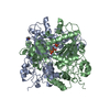
Entry Database : PDB / ID : 7ej3Title UTP cyclohydrolase GTP cyclohydrolase II Keywords / / / Function / homology Function Domain/homology Component
/ / / / / Biological species Rhodococcus wratislaviensis (bacteria)Method / / / Resolution : 1.6 Å Authors Zhang, H. / Zhang, Y. / Yuchi, Z. Funding support Organization Grant number Country National Science Foundation (NSF, China) 31870049
Journal : Acs Catalysis / Year : 2021Title : Structural and biochemical investigation of UTP cyclohydrolaseAuthors : Zhang, H. / Wei, Y. / Lin, L. / Liu, J. / Chu, R. / Cao, P. / Ang, E.L. / Zhao, H. / Yuchi, Z. / Zhang, Y. History Deposition Apr 1, 2021 Deposition site / Processing site Revision 1.0 Sep 21, 2022 Provider / Type Revision 1.1 May 29, 2024 Group / Refinement descriptionCategory / chem_comp_bond / struct_ncs_dom_limItem _struct_ncs_dom_lim.beg_auth_comp_id / _struct_ncs_dom_lim.beg_label_asym_id ... _struct_ncs_dom_lim.beg_auth_comp_id / _struct_ncs_dom_lim.beg_label_asym_id / _struct_ncs_dom_lim.beg_label_comp_id / _struct_ncs_dom_lim.beg_label_seq_id / _struct_ncs_dom_lim.end_auth_comp_id / _struct_ncs_dom_lim.end_label_asym_id / _struct_ncs_dom_lim.end_label_comp_id / _struct_ncs_dom_lim.end_label_seq_id
Show all Show less
 Movie
Movie Controller


 Open data
 Basic information
 Components
 Keywords
 Function and homology information
 Rhodococcus wratislaviensis (bacteria)
 X-RAY DIFFRACTION /
  Authors
 China, 1items
  Citation
 Structure visualization
 Downloads & links
 Other downloads
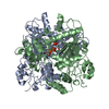
 PDBj
PDBj

 Assembly