 Movie
Movie Controller


+ Open data

- Basic information
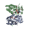
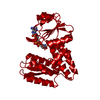
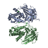
| Entry | Database: PDB / ID: 7ehm | ||||||
|---|---|---|---|---|---|---|---|
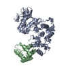
| Title | Human MTHFD2 in complex with compound 21 and 15 | ||||||
 Components Components | Bifunctional methylenetetrahydrofolate dehydrogenase/cyclohydrolase, mitochondrial | ||||||
 Keywords Keywords | OXIDOREDUCTASE / MTHFD2 / methylenetetrahydrofolate dehydrogenase 2 / 1C metabolism / mitocondria | ||||||
| Function / homology |  Function and homology information Function and homology informationmethylenetetrahydrofolate dehydrogenase (NAD+) / methylenetetrahydrofolate dehydrogenase (NAD+) activity / formate biosynthetic process / methenyltetrahydrofolate cyclohydrolase / methenyltetrahydrofolate cyclohydrolase activity / methylenetetrahydrofolate dehydrogenase (NADP+) activity / Metabolism of folate and pterines / tetrahydrofolate metabolic process / tetrahydrofolate interconversion / phosphate ion binding ...methylenetetrahydrofolate dehydrogenase (NAD+) / methylenetetrahydrofolate dehydrogenase (NAD+) activity / formate biosynthetic process / methenyltetrahydrofolate cyclohydrolase / methenyltetrahydrofolate cyclohydrolase activity / methylenetetrahydrofolate dehydrogenase (NADP+) activity / Metabolism of folate and pterines / tetrahydrofolate metabolic process / tetrahydrofolate interconversion / phosphate ion binding / folic acid metabolic process / mitochondrial matrix / magnesium ion binding / mitochondrion / : Similarity search - Function | ||||||
| Biological species |  Homo sapiens (human) Homo sapiens (human) | ||||||
| Method |  X-RAY DIFFRACTION / SYNCHROTRON / MOLECULAR REPLACEMENT / molecular replacement / Resolution: 2.13 Å X-RAY DIFFRACTION / SYNCHROTRON / MOLECULAR REPLACEMENT / molecular replacement / Resolution: 2.13 Å | ||||||
 Authors Authors | Lee, L.C. / Peng, Y.H. / Wu, S.Y. | ||||||
 Citation Citation | Journal: J.Med.Chem. / Year: 2021 Title: Xanthine Derivatives Reveal an Allosteric Binding Site in Methylenetetrahydrofolate Dehydrogenase 2 (MTHFD2). Authors: Lee, L.C. / Peng, Y.H. / Chang, H.H. / Hsu, T. / Lu, C.T. / Huang, C.H. / Hsueh, C.C. / Kung, F.C. / Kuo, C.C. / Jiaang, W.T. / Wu, S.Y. | ||||||
| History |
|
- Structure visualization
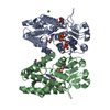
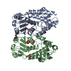
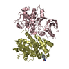
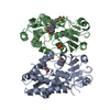
| Structure viewer | Molecule: MolmilJmol/JSmol |
|---|
- Downloads & links
-Download
| PDBx/mmCIF format | 7ehm.cif.gz | 129.9 KB | Display | PDBx/mmCIF format |
|---|---|---|---|---|
| PDB format | pdb7ehm.ent.gz | 98 KB | Display | PDB format |
| PDBx/mmJSON format | 7ehm.json.gz | Tree view | PDBx/mmJSON format | |
| Others |  Other downloads Other downloads |
-Validation report
| Arichive directory | https://data.pdbj.org/pub/pdb/validation_reports/eh/7ehmftp://data.pdbj.org/pub/pdb/validation_reports/eh/7ehm | HTTPS FTP |
|---|
-Related structure data
| Related structure data | 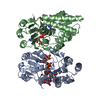 7ehjSC 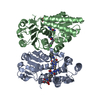 7ehnC 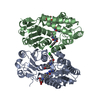 7ehvC S: Starting model for refinement C: citing same article ( |
|---|---|
| Similar structure data |


-Links
 PDBj
PDBj
- Assembly
| Deposited unit | 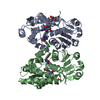
| ||||||||
|---|---|---|---|---|---|---|---|---|---|
| 1 |
| ||||||||
| Unit cell |
|
-Components
| #1: Protein | Mass: 34183.555 Da / Num. of mol.: 2 Source method: isolated from a genetically manipulated source Source: (gene. exp.) Homo sapiens (human) / Gene: MTHFD2, NMDMC / Plasmid: pET14b / Production host:  References: UniProt: P13995, methylenetetrahydrofolate dehydrogenase (NAD+), methenyltetrahydrofolate cyclohydrolase #2: Chemical | 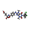  Mass: 603.411 Da / Num. of mol.: 2 / Source method: obtained synthetically / Formula: C26H24Cl2N6O7 / Feature type: SUBJECT OF INVESTIGATION Mass: 603.411 Da / Num. of mol.: 2 / Source method: obtained synthetically / Formula: C26H24Cl2N6O7 / Feature type: SUBJECT OF INVESTIGATION#3: Chemical | 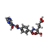  Mass: 418.361 Da / Num. of mol.: 2 / Source method: obtained synthetically / Formula: C17H18N6O7 Mass: 418.361 Da / Num. of mol.: 2 / Source method: obtained synthetically / Formula: C17H18N6O7#4: Water | ChemComp-HOH / |  Mass: 18.015 Da / Num. of mol.: 197 / Source method: isolated from a natural source / Formula: H2O Mass: 18.015 Da / Num. of mol.: 197 / Source method: isolated from a natural source / Formula: H2OHas ligand of interest | Y | |
|---|
-Experimental details
-Experiment
| Experiment | Method: X-RAY DIFFRACTION / Number of used crystals: 1 |
|---|
- Sample preparation
| Crystal | Density Matthews: 2.95 Å3/Da / Density % sol: 58.26 % |
|---|---|
| Crystal grow | Temperature: 291.15 K / Method: vapor diffusion, hanging drop / pH: 7.1 Details: isopropanol, bis-Tris pH 7.1, PEG 200, PEG 3350, glycerol |
-Data collection
| Diffraction | Mean temperature: 100 K / Serial crystal experiment: N | |||||||||||||||||||||||||||||||||||||||||||||||||||||||||||||||||||||||||||||||||||||||||||||||||||
|---|---|---|---|---|---|---|---|---|---|---|---|---|---|---|---|---|---|---|---|---|---|---|---|---|---|---|---|---|---|---|---|---|---|---|---|---|---|---|---|---|---|---|---|---|---|---|---|---|---|---|---|---|---|---|---|---|---|---|---|---|---|---|---|---|---|---|---|---|---|---|---|---|---|---|---|---|---|---|---|---|---|---|---|---|---|---|---|---|---|---|---|---|---|---|---|---|---|---|---|---|
| Diffraction source | Source: SYNCHROTRON / Site: NSRRC  / Beamline: TPS 05A / Wavelength: 1 Å / Beamline: TPS 05A / Wavelength: 1 Å | |||||||||||||||||||||||||||||||||||||||||||||||||||||||||||||||||||||||||||||||||||||||||||||||||||
| Detector | Type: RAYONIX MX300-HS / Detector: CCD / Date: Mar 6, 2019 / Details: A Pair of K-B Focusing Mirrors | |||||||||||||||||||||||||||||||||||||||||||||||||||||||||||||||||||||||||||||||||||||||||||||||||||
| Radiation | Monochromator: LN2-cooled, Fixed-Exit Double Crystal Monochromator Protocol: SINGLE WAVELENGTH / Monochromatic (M) / Laue (L): M / Scattering type: x-ray | |||||||||||||||||||||||||||||||||||||||||||||||||||||||||||||||||||||||||||||||||||||||||||||||||||
| Radiation wavelength | Wavelength: 1 Å / Relative weight: 1 | |||||||||||||||||||||||||||||||||||||||||||||||||||||||||||||||||||||||||||||||||||||||||||||||||||
| Reflection | Resolution: 2.13→30 Å / Num. obs: 47198 / % possible obs: 99.9 % / Redundancy: 4.4 % / Rmerge(I) obs: 0.061 / Rpim(I) all: 0.031 / Rrim(I) all: 0.069 / Χ2: 1.059 / Net I/σ(I): 9.9 / Num. measured all: 209917 | |||||||||||||||||||||||||||||||||||||||||||||||||||||||||||||||||||||||||||||||||||||||||||||||||||
| Reflection shell | Diffraction-ID: 1
|
-Phasing
| Phasing | Method: molecular replacement | ||||||
|---|---|---|---|---|---|---|---|
| Phasing MR | R rigid body: 0.403
|
- Processing
| Software |
| |||||||||||||||||||||||||||||||||||||||||||||||||||||||||||||||||||||||||||||||||||||||||||||||||||||||||||||||||||||||
|---|---|---|---|---|---|---|---|---|---|---|---|---|---|---|---|---|---|---|---|---|---|---|---|---|---|---|---|---|---|---|---|---|---|---|---|---|---|---|---|---|---|---|---|---|---|---|---|---|---|---|---|---|---|---|---|---|---|---|---|---|---|---|---|---|---|---|---|---|---|---|---|---|---|---|---|---|---|---|---|---|---|---|---|---|---|---|---|---|---|---|---|---|---|---|---|---|---|---|---|---|---|---|---|---|---|---|---|---|---|---|---|---|---|---|---|---|---|---|---|---|
| Refinement | Method to determine structure: MOLECULAR REPLACEMENT Starting model: 7EHJ Resolution: 2.13→24.92 Å / SU ML: 0.23 / Cross valid method: THROUGHOUT / σ(F): 1.34 / Phase error: 24.29 / Stereochemistry target values: ML
| |||||||||||||||||||||||||||||||||||||||||||||||||||||||||||||||||||||||||||||||||||||||||||||||||||||||||||||||||||||||
| Solvent computation | Shrinkage radii: 0.9 Å / VDW probe radii: 1.11 Å / Solvent model: FLAT BULK SOLVENT MODEL | |||||||||||||||||||||||||||||||||||||||||||||||||||||||||||||||||||||||||||||||||||||||||||||||||||||||||||||||||||||||
| Displacement parameters | Biso max: 89.62 Å2 / Biso mean: 45.1056 Å2 / Biso min: 23.92 Å2 | |||||||||||||||||||||||||||||||||||||||||||||||||||||||||||||||||||||||||||||||||||||||||||||||||||||||||||||||||||||||
| Refinement step | Cycle: final / Resolution: 2.13→24.92 Å
| |||||||||||||||||||||||||||||||||||||||||||||||||||||||||||||||||||||||||||||||||||||||||||||||||||||||||||||||||||||||
| LS refinement shell | Refine-ID: X-RAY DIFFRACTION / Rfactor Rfree error: 0 / Total num. of bins used: 16
|
