 Movie
Movie Controller

[English] 日本語
 Yorodumi
- PDB-7egs: The crystal structure of lobe domain of E. coli RNA polymerase co... -
+ Open data

- Basic information
| Entry | Database: PDB / ID: 7egs | ||||||
|---|---|---|---|---|---|---|---|
| Title | The crystal structure of lobe domain of E. coli RNA polymerase complexed with the C-terminal domain of UvrD | ||||||
 Components Components |
| ||||||
 Keywords Keywords | TRANSCRIPTION / TCR / Escherichia Coli / UvrD / RNA polymerase / DNA repair | ||||||
| Function / homology |  Function and homology information Function and homology informationrolling circle DNA replication / single-stranded DNA-dependent ATP-dependent DNA helicase complex / mismatch repair involved in maintenance of fidelity involved in DNA-dependent DNA replication / DNA helicase complex / DNA translocase activity / single-stranded DNA helicase activity / SOS response / recombinational repair / 3'-5' DNA helicase activity / submerged biofilm formation ...rolling circle DNA replication / single-stranded DNA-dependent ATP-dependent DNA helicase complex / mismatch repair involved in maintenance of fidelity involved in DNA-dependent DNA replication / DNA helicase complex / DNA translocase activity / single-stranded DNA helicase activity / SOS response / recombinational repair / 3'-5' DNA helicase activity / submerged biofilm formation / DNA 3'-5' helicase / cellular response to cell envelope stress / regulation of DNA-templated transcription initiation / replication fork processing / bacterial-type flagellum assembly / cytosolic DNA-directed RNA polymerase complex / bacterial-type flagellum-dependent cell motility / nitrate assimilation / mismatch repair / DNA helicase activity / regulation of DNA-templated transcription elongation / transcription elongation factor complex / transcription antitermination / nucleotide-excision repair / cell motility / DNA-templated transcription initiation / response to radiation / ribonucleoside binding / DNA-directed RNA polymerase / DNA-directed RNA polymerase activity / response to heat / intracellular iron ion homeostasis / response to antibiotic / protein homodimerization activity / ATP hydrolysis activity / DNA binding / ATP binding / membrane / cytoplasm / cytosol Similarity search - Function | ||||||
| Biological species |  | ||||||
| Method |  X-RAY DIFFRACTION / SYNCHROTRON / MOLECULAR REPLACEMENT / Resolution: 1.7 Å X-RAY DIFFRACTION / SYNCHROTRON / MOLECULAR REPLACEMENT / Resolution: 1.7 Å | ||||||
 Authors Authors | Zheng, F. / Shen, L. / Li, L. / Zhang, Y. | ||||||
| Funding support |  China, 1items China, 1items
| ||||||
 Citation Citation | Journal: Nature / Year: 2022 Title: Crucial role and mechanism of transcription-coupled DNA repair in bacteria. Authors: Bharati, B.K. / Gowder, M. / Zheng, F. / Alzoubi, K. / Svetlov, V. / Kamarthapu, V. / Weaver, J.W. / Epshtein, V. / Vasilyev, N. / Shen, L. / Zhang, Y. / Nudler, E. | ||||||
| History |
|
- Structure visualization
| Structure viewer | Molecule: MolmilJmol/JSmol |
|---|
- Downloads & links
-Download
| PDBx/mmCIF format | 7egs.cif.gz | 89.2 KB | Display | PDBx/mmCIF format |
|---|---|---|---|---|
| PDB format | pdb7egs.ent.gz | 63.6 KB | Display | PDB format |
| PDBx/mmJSON format | 7egs.json.gz | Tree view | PDBx/mmJSON format | |
| Others |  Other downloads Other downloads |
-Validation report
| Arichive directory | https://data.pdbj.org/pub/pdb/validation_reports/eg/7egsftp://data.pdbj.org/pub/pdb/validation_reports/eg/7egs | HTTPS FTP |
|---|
-Related structure data
| Related structure data | 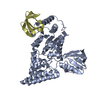 7egtC 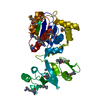 3ltiS S: Starting model for refinement C: citing same article ( |
|---|---|
| Similar structure data |
-Links
 PDBj
PDBj


- Assembly
| Deposited unit | 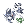
| ||||||||
|---|---|---|---|---|---|---|---|---|---|
| 1 |
| ||||||||
| Unit cell |
|
-Components
| #1: Protein | Mass: 33931.730 Da / Num. of mol.: 1 Source method: isolated from a genetically manipulated source Source: (gene. exp.) Strain: K12 Gene: rpoB, groN, nitB, rif, ron, stl, stv, tabD, b3987, JW3950 Production host: |
|---|---|
| #2: Protein | Mass: 7729.751 Da / Num. of mol.: 1 Source method: isolated from a genetically manipulated source Source: (gene. exp.) Strain: K12 / Gene: uvrD, mutU, pdeB, rad, recL, b3813, JW3786 / Production host: |
| #3: Chemical | ChemComp-GOL /   Mass: 92.094 Da / Num. of mol.: 1 / Source method: obtained synthetically / Formula: C3H8O3 Mass: 92.094 Da / Num. of mol.: 1 / Source method: obtained synthetically / Formula: C3H8O3 |
| #4: Water | ChemComp-HOH /  Mass: 18.015 Da / Num. of mol.: 161 / Source method: isolated from a natural source / Formula: H2O Mass: 18.015 Da / Num. of mol.: 161 / Source method: isolated from a natural source / Formula: H2O |
| Has ligand of interest | N |
-Experimental details
-Experiment
| Experiment | Method: X-RAY DIFFRACTION / Number of used crystals: 1 |
|---|
- Sample preparation
| Crystal | Density Matthews: 2.58 Å3/Da / Density % sol: 52.36 % |
|---|---|
| Crystal grow | Temperature: 295 K / Method: vapor diffusion, hanging drop Details: 0.1 M HEPES pH 7.0, 10% w/v polyethylene glycol 4000, 10 % (v/v) isopropanol |
-Data collection
| Diffraction | Mean temperature: 100 K / Serial crystal experiment: N | |||||||||||||||||||||||||||||||||||||||||||||||||||||||||||||||||||||||||||||||||||||||||||||||||||||||||||||||||||||||||||||||||||||||||||||||||||||||||||||||||||||||||||||||||||||||||||||
|---|---|---|---|---|---|---|---|---|---|---|---|---|---|---|---|---|---|---|---|---|---|---|---|---|---|---|---|---|---|---|---|---|---|---|---|---|---|---|---|---|---|---|---|---|---|---|---|---|---|---|---|---|---|---|---|---|---|---|---|---|---|---|---|---|---|---|---|---|---|---|---|---|---|---|---|---|---|---|---|---|---|---|---|---|---|---|---|---|---|---|---|---|---|---|---|---|---|---|---|---|---|---|---|---|---|---|---|---|---|---|---|---|---|---|---|---|---|---|---|---|---|---|---|---|---|---|---|---|---|---|---|---|---|---|---|---|---|---|---|---|---|---|---|---|---|---|---|---|---|---|---|---|---|---|---|---|---|---|---|---|---|---|---|---|---|---|---|---|---|---|---|---|---|---|---|---|---|---|---|---|---|---|---|---|---|---|---|---|---|---|
| Diffraction source | Source: SYNCHROTRON / Site: SSRF / Beamline: BL17U1 / Wavelength: 0.97853 Å | |||||||||||||||||||||||||||||||||||||||||||||||||||||||||||||||||||||||||||||||||||||||||||||||||||||||||||||||||||||||||||||||||||||||||||||||||||||||||||||||||||||||||||||||||||||||||||||
| Detector | Type: ADSC QUANTUM 315r / Detector: CCD / Date: May 30, 2016 | |||||||||||||||||||||||||||||||||||||||||||||||||||||||||||||||||||||||||||||||||||||||||||||||||||||||||||||||||||||||||||||||||||||||||||||||||||||||||||||||||||||||||||||||||||||||||||||
| Radiation | Protocol: SINGLE WAVELENGTH / Monochromatic (M) / Laue (L): M / Scattering type: x-ray | |||||||||||||||||||||||||||||||||||||||||||||||||||||||||||||||||||||||||||||||||||||||||||||||||||||||||||||||||||||||||||||||||||||||||||||||||||||||||||||||||||||||||||||||||||||||||||||
| Radiation wavelength | Wavelength: 0.97853 Å / Relative weight: 1 | |||||||||||||||||||||||||||||||||||||||||||||||||||||||||||||||||||||||||||||||||||||||||||||||||||||||||||||||||||||||||||||||||||||||||||||||||||||||||||||||||||||||||||||||||||||||||||||
| Reflection | Resolution: 1.7→40 Å / Num. obs: 46583 / % possible obs: 98.9 % / Redundancy: 3.3 % / Biso Wilson estimate: 29.57 Å2 / Rmerge(I) obs: 0.065 / Rpim(I) all: 0.041 / Rrim(I) all: 0.077 / Χ2: 1.137 / Net I/av σ(I): 16.3 / Net I/σ(I): 16.3 | |||||||||||||||||||||||||||||||||||||||||||||||||||||||||||||||||||||||||||||||||||||||||||||||||||||||||||||||||||||||||||||||||||||||||||||||||||||||||||||||||||||||||||||||||||||||||||||
| Reflection shell | Diffraction-ID: 1
|
- Processing
| Software |
| ||||||||||||||||||||||||||||||||||||||||||||||||||||||||||||||||||||||||||||||||||||||||||||||||||||||||||||
|---|---|---|---|---|---|---|---|---|---|---|---|---|---|---|---|---|---|---|---|---|---|---|---|---|---|---|---|---|---|---|---|---|---|---|---|---|---|---|---|---|---|---|---|---|---|---|---|---|---|---|---|---|---|---|---|---|---|---|---|---|---|---|---|---|---|---|---|---|---|---|---|---|---|---|---|---|---|---|---|---|---|---|---|---|---|---|---|---|---|---|---|---|---|---|---|---|---|---|---|---|---|---|---|---|---|---|---|---|---|
| Refinement | Method to determine structure: MOLECULAR REPLACEMENT Starting model: 3LTI Resolution: 1.7→37.807 Å / SU ML: 0.22 / Cross valid method: THROUGHOUT / σ(F): 1.38 / Phase error: 25.9 / Stereochemistry target values: ML
| ||||||||||||||||||||||||||||||||||||||||||||||||||||||||||||||||||||||||||||||||||||||||||||||||||||||||||||
| Solvent computation | Shrinkage radii: 0.9 Å / VDW probe radii: 1.11 Å / Solvent model: FLAT BULK SOLVENT MODEL | ||||||||||||||||||||||||||||||||||||||||||||||||||||||||||||||||||||||||||||||||||||||||||||||||||||||||||||
| Displacement parameters | Biso max: 84.83 Å2 / Biso mean: 38.6115 Å2 / Biso min: 19.14 Å2 | ||||||||||||||||||||||||||||||||||||||||||||||||||||||||||||||||||||||||||||||||||||||||||||||||||||||||||||
| Refinement step | Cycle: final / Resolution: 1.7→37.807 Å
| ||||||||||||||||||||||||||||||||||||||||||||||||||||||||||||||||||||||||||||||||||||||||||||||||||||||||||||
| Refine LS restraints |
| ||||||||||||||||||||||||||||||||||||||||||||||||||||||||||||||||||||||||||||||||||||||||||||||||||||||||||||
| LS refinement shell | Refine-ID: X-RAY DIFFRACTION / Rfactor Rfree error: 0
|
