 Movie
Movie Controller

[English] 日本語
 Yorodumi
- PDB-7eg5: FMN-bound form of YviC from Lactococcus lactis subsp. lactis Il1403 -
+ Open data

- Basic information
| Entry | Database: PDB / ID: 7eg5 | ||||||
|---|---|---|---|---|---|---|---|
| Title | FMN-bound form of YviC from Lactococcus lactis subsp. lactis Il1403 | ||||||
 Components Components | FMN-binding protein | ||||||
 Keywords Keywords | FLAVOPROTEIN / fmn binding | ||||||
| Function / homology | Pyridoxamine 5'-phosphate oxidase, putative / Pyridoxamine 5'-phosphate oxidase / FMN-binding split barrel / nucleotide binding / FLAVIN MONONUCLEOTIDE / FMN-binding protein Function and homology information Function and homology information | ||||||
| Biological species |  Lactococcus lactis subsp. lactis Il1403 (lactic acid bacteria) Lactococcus lactis subsp. lactis Il1403 (lactic acid bacteria) | ||||||
| Method |  X-RAY DIFFRACTION / MOLECULAR REPLACEMENT / Resolution: 1.96 Å X-RAY DIFFRACTION / MOLECULAR REPLACEMENT / Resolution: 1.96 Å | ||||||
 Authors Authors | Sugiura, N. / Nakanishi, T. / Kitamura, M. | ||||||
 Citation Citation | Journal: To Be Published Title: FMN-bound form of YviC from Lactococcus lactis subsp. lactis Il1403 Authors: Sugiura, N. / Nakanishi, T. / Kitamura, M. | ||||||
| History |
|
- Structure visualization
| Structure viewer | Molecule: MolmilJmol/JSmol |
|---|
- Downloads & links
-Download
| PDBx/mmCIF format | 7eg5.cif.gz | 63.6 KB | Display | PDBx/mmCIF format |
|---|---|---|---|---|
| PDB format | pdb7eg5.ent.gz | 46.8 KB | Display | PDB format |
| PDBx/mmJSON format | 7eg5.json.gz | Tree view | PDBx/mmJSON format | |
| Others |  Other downloads Other downloads |
-Validation report
| Summary document | 7eg5_validation.pdf.gz | 1.1 MB | Display | wwPDB validaton report |
|---|---|---|---|---|
| Full document | 7eg5_full_validation.pdf.gz | 1.1 MB | Display | |
| Data in XML | 7eg5_validation.xml.gz | 12 KB | Display | |
| Data in CIF | 7eg5_validation.cif.gz | 15 KB | Display | |
| Arichive directory | https://data.pdbj.org/pub/pdb/validation_reports/eg/7eg5ftp://data.pdbj.org/pub/pdb/validation_reports/eg/7eg5 | HTTPS FTP |
-Related structure data
| Related structure data | 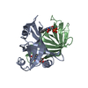 1flmS S: Starting model for refinement |
|---|---|
| Similar structure data |
-Links
 PDBj
PDBj- Assembly
| Deposited unit | 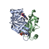
| ||||||||
|---|---|---|---|---|---|---|---|---|---|
| 1 |
| ||||||||
| Unit cell |
|
-Components
| #1: Protein | Mass: 14147.332 Da / Num. of mol.: 2 Source method: isolated from a genetically manipulated source Source: (gene. exp.) Lactococcus lactis subsp. lactis Il1403 (lactic acid bacteria)Strain: IL1403 / Gene: yviC, L164222 / Production host: #2: Chemical |   Mass: 456.344 Da / Num. of mol.: 2 / Source method: obtained synthetically / Formula: C17H21N4O9P Mass: 456.344 Da / Num. of mol.: 2 / Source method: obtained synthetically / Formula: C17H21N4O9P#3: Water | ChemComp-HOH / |  Mass: 18.015 Da / Num. of mol.: 16 / Source method: isolated from a natural source / Formula: H2O Mass: 18.015 Da / Num. of mol.: 16 / Source method: isolated from a natural source / Formula: H2OHas ligand of interest | Y | |
|---|
-Experimental details
-Experiment
| Experiment | Method: X-RAY DIFFRACTION / Number of used crystals: 1 |
|---|
- Sample preparation
| Crystal | Density Matthews: 2.47 Å3/Da / Density % sol: 50.2 % Description: THE ENTRY CONTAINS FRIEDEL PAIRS IN I/F_PLUS/MINUS COLUMNS. |
|---|---|
| Crystal grow | Temperature: 293 K / Method: vapor diffusion, hanging drop / Details: 0.1 M HEPES (pH 7.62), 1.7 M Ammonium sulfate |
-Data collection
| Diffraction | Mean temperature: 100 K / Serial crystal experiment: N |
|---|---|
| Diffraction source | Source: ROTATING ANODE / Type: RIGAKU FR-X / Wavelength: 1.5418 Å |
| Detector | Type: RIGAKU RAXIS VII / Detector: IMAGE PLATE / Date: Oct 29, 2020 |
| Radiation | Protocol: SINGLE WAVELENGTH / Monochromatic (M) / Laue (L): M / Scattering type: x-ray |
| Radiation wavelength | Wavelength: 1.5418 Å / Relative weight: 1 |
| Reflection | Resolution: 1.96→19.93 Å / Num. obs: 20447 / % possible obs: 99.3 % / Redundancy: 8.2 % / Rmerge(I) obs: 0.07 / Net I/σ(I): 16.1 |
| Reflection shell | Resolution: 1.96→2.07 Å / Redundancy: 7.6 % / Rmerge(I) obs: 0.378 / % possible all: 98.7 |
- Processing
| Software |
| ||||||||||||||||||||||||||||||||||||||||||||||||||||||||||||||||||||||||||||||||||||||||||||||||||||||||||||||||||||||||||||||||||||||||||||||||||||||||||||||||||||||||||||||||||||||
|---|---|---|---|---|---|---|---|---|---|---|---|---|---|---|---|---|---|---|---|---|---|---|---|---|---|---|---|---|---|---|---|---|---|---|---|---|---|---|---|---|---|---|---|---|---|---|---|---|---|---|---|---|---|---|---|---|---|---|---|---|---|---|---|---|---|---|---|---|---|---|---|---|---|---|---|---|---|---|---|---|---|---|---|---|---|---|---|---|---|---|---|---|---|---|---|---|---|---|---|---|---|---|---|---|---|---|---|---|---|---|---|---|---|---|---|---|---|---|---|---|---|---|---|---|---|---|---|---|---|---|---|---|---|---|---|---|---|---|---|---|---|---|---|---|---|---|---|---|---|---|---|---|---|---|---|---|---|---|---|---|---|---|---|---|---|---|---|---|---|---|---|---|---|---|---|---|---|---|---|---|---|---|---|
| Refinement | Method to determine structure: MOLECULAR REPLACEMENT Starting model: 1FLM Resolution: 1.96→19.93 Å / Cor.coef. Fo:Fc: 0.96 / Cor.coef. Fo:Fc free: 0.947 / SU B: 3.762 / SU ML: 0.108 / Cross valid method: THROUGHOUT / σ(F): 0 / ESU R: 0.171 / ESU R Free: 0.149 / Stereochemistry target values: MAXIMUM LIKELIHOOD / Details: HYDROGENS HAVE BEEN ADDED IN THE RIDING
| ||||||||||||||||||||||||||||||||||||||||||||||||||||||||||||||||||||||||||||||||||||||||||||||||||||||||||||||||||||||||||||||||||||||||||||||||||||||||||||||||||||||||||||||||||||||
| Solvent computation | Ion probe radii: 0.8 Å / Shrinkage radii: 0.8 Å / VDW probe radii: 1.2 Å / Solvent model: MASK | ||||||||||||||||||||||||||||||||||||||||||||||||||||||||||||||||||||||||||||||||||||||||||||||||||||||||||||||||||||||||||||||||||||||||||||||||||||||||||||||||||||||||||||||||||||||
| Displacement parameters | Biso mean: 41.74 Å2
| ||||||||||||||||||||||||||||||||||||||||||||||||||||||||||||||||||||||||||||||||||||||||||||||||||||||||||||||||||||||||||||||||||||||||||||||||||||||||||||||||||||||||||||||||||||||
| Refinement step | Cycle: LAST / Resolution: 1.96→19.93 Å
| ||||||||||||||||||||||||||||||||||||||||||||||||||||||||||||||||||||||||||||||||||||||||||||||||||||||||||||||||||||||||||||||||||||||||||||||||||||||||||||||||||||||||||||||||||||||
| Refine LS restraints |
|