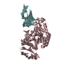
- PDB-7ef9: Crystal structure of mouse MUTYH in complex with DNA containing A... -
+
Open data
ID or keywords:
Loading...
-
Basic information
Entry
Database: PDB / ID: 7ef9
Title
Crystal structure of mouse MUTYH in complex with DNA containing AP site analogue:8-oxoG (Form II)
Components
Adenine DNA glycosylase
DNA (5'-D(*AP*TP*GP*AP*GP*AP*CP*(8OG)P*GP*GP*GP*AP*CP*T)-3')
DNA (5'-D(*TP*AP*GP*TP*CP*CP*CP*(3DR)P*GP*TP*CP*TP*C)-3')
Keywords
DNA BINDING PROTEIN/DNA / DNA repair / DNA BINDING PROTEIN-DNA complex
Function / homology
Function and homology information
Displacement of DNA glycosylase by APEX1 / adenine glycosylase / adenine/guanine mispair binding / Cleavage of the damaged purine / MutSalpha complex binding / DNA N-glycosylase activity / 8-oxo-7,8-dihydroguanine DNA N-glycosylase activity / purine-specific mismatch base pair DNA N-glycosylase activity / negative regulation of necroptotic process / oxidized purine DNA binding ...Displacement of DNA glycosylase by APEX1 / adenine glycosylase / adenine/guanine mispair binding / Cleavage of the damaged purine / MutSalpha complex binding / DNA N-glycosylase activity / 8-oxo-7,8-dihydroguanine DNA N-glycosylase activity / purine-specific mismatch base pair DNA N-glycosylase activity / negative regulation of necroptotic process / oxidized purine DNA binding / mismatch repair / base-excision repair / 4 iron, 4 sulfur cluster binding / response to oxidative stress / DNA repair / mitochondrion / nucleoplasm / metal ion binding / nucleus Similarity search - Function
Endonuclease III, iron-sulphur binding site / Endonuclease III-like, conserved site-2 / Endonuclease III iron-sulfur binding region signature. / Endonuclease III family signature. / Iron-sulfur binding domain of endonuclease III / Adenine/Thymine-DNA glycosylase / MutY, C-terminal / NUDIX domain / Helix-hairpin-helix motif / Endonuclease III-like, iron-sulphur cluster loop motif ...Endonuclease III, iron-sulphur binding site / Endonuclease III-like, conserved site-2 / Endonuclease III iron-sulfur binding region signature. / Endonuclease III family signature. / Iron-sulfur binding domain of endonuclease III / Adenine/Thymine-DNA glycosylase / MutY, C-terminal / NUDIX domain / Helix-hairpin-helix motif / Endonuclease III-like, iron-sulphur cluster loop motif / FES / Helix-hairpin-helix motif / HhH-GPD superfamily base excision DNA repair protein / Helix-hairpin-helix, base-excision DNA repair, C-terminal / HhH-GPD domain / endonuclease III / DNA glycosylase / Nucleoside Triphosphate Pyrophosphohydrolase / Nucleoside Triphosphate Pyrophosphohydrolase / Nudix hydrolase domain profile. / NUDIX hydrolase domain / NUDIX hydrolase-like domain superfamily / Alpha-Beta Complex / Alpha Beta Similarity search - Domain/homology
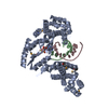
A: Adenine DNA glycosylase B: DNA (5'-D(*AP*TP*GP*AP*GP*AP*CP*(8OG)P*GP*GP*GP*AP*CP*T)-3') C: DNA (5'-D(*TP*AP*GP*TP*CP*CP*CP*(3DR)P*GP*TP*CP*TP*C)-3') hetero molecules
In the structure databanks used in Yorodumi, some data are registered as the other names, "COVID-19 virus" and "2019-nCoV". Here are the details of the virus and the list of structure data.
Jan 31, 2019. EMDB accession codes are about to change! (news from PDBe EMDB page)
EMDB accession codes are about to change! (news from PDBe EMDB page)
The allocation of 4 digits for EMDB accession codes will soon come to an end. Whilst these codes will remain in use, new EMDB accession codes will include an additional digit and will expand incrementally as the available range of codes is exhausted. The current 4-digit format prefixed with “EMD-” (i.e. EMD-XXXX) will advance to a 5-digit format (i.e. EMD-XXXXX), and so on. It is currently estimated that the 4-digit codes will be depleted around Spring 2019, at which point the 5-digit format will come into force.
The EM Navigator/Yorodumi systems omit the EMD- prefix.
Related info.:Q: What is EMD? / ID/Accession-code notation in Yorodumi/EM Navigator
Yorodumi is a browser for structure data from EMDB, PDB, SASBDB, etc.
This page is also the successor to EM Navigator detail page, and also detail information page/front-end page for Omokage search.
The word "yorodu" (or yorozu) is an old Japanese word meaning "ten thousand". "mi" (miru) is to see.
Related info.:EMDB / PDB / SASBDB / Comparison of 3 databanks / Yorodumi Search / Aug 31, 2016. New EM Navigator & Yorodumi / Yorodumi Papers / Jmol/JSmol / Function and homology information / Changes in new EM Navigator and Yorodumi
 Movie
Movie Controller

 Yorodumi
 Open data
 Basic information
 Components
 Keywords
 Function and homology information
 Homo sapiens (human)
 X-RAY DIFFRACTION /
  Authors
 Japan, 2items
  Citation
 Structure visualization
 Downloads & links
 Other downloads
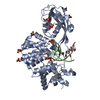


 PDBj
PDBj


 Assembly
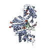


 Mass: 351.640 Da / Num. of mol.: 1 / Source method: obtained synthetically / Formula: Fe4S4
 Mass: 65.409 Da / Num. of mol.: 1 / Source method: obtained synthetically / Formula: Zn
 Mass: 96.063 Da / Num. of mol.: 6 / Source method: obtained synthetically / Formula: SO4
 Mass: 92.094 Da / Num. of mol.: 1 / Source method: obtained synthetically / Formula: C3H8O3
 Sample preparation
 Processing