 Movie
Movie Controller

[English] 日本語
 Yorodumi
- PDB-7ecd: Crystal structure of Tam41 from Firmicutes bacterium, complex wit... -
+ Open data

- Basic information
| Entry | Database: PDB / ID: 7ecd | |||||||||||||||
|---|---|---|---|---|---|---|---|---|---|---|---|---|---|---|---|---|
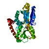
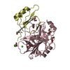
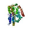
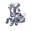
| Title | Crystal structure of Tam41 from Firmicutes bacterium, complex with CTP-Mg | |||||||||||||||
 Components Components | Phosphatidate cytidylyltransferase | |||||||||||||||
 Keywords Keywords | TRANSFERASE / CDP-DAG synthase / phosphatidic acid / cytidine-diphosphate diacylglycerol / Mitochondrial matrix | |||||||||||||||
| Function / homology |  Function and homology information Function and homology informationphosphatidate cytidylyltransferase / phosphatidate cytidylyltransferase activity / CDP-diacylglycerol biosynthetic process / cardiolipin biosynthetic process / membrane Similarity search - Function | |||||||||||||||
| Biological species |  Firmicutes bacterium CAG:884 (bacteria) Firmicutes bacterium CAG:884 (bacteria) | |||||||||||||||
| Method |  X-RAY DIFFRACTION / SYNCHROTRON / SAD / Resolution: 2.6 Å X-RAY DIFFRACTION / SYNCHROTRON / SAD / Resolution: 2.6 Å | |||||||||||||||
 Authors Authors | Kimura, K. / Kawai, F. / Kubota-Kawai, H. / Watanabe, Y. / Tamura, Y. | |||||||||||||||
| Funding support |  Japan, 4items Japan, 4items
| |||||||||||||||
 Citation Citation | Journal: J.Biochem. / Year: 2022 Title: Crystal structure of Tam41 cytidine diphosphate diacylglycerol synthase from a Firmicutes bacterium. Authors: Kimura, K. / Kawai, F. / Kubota-Kawai, H. / Watanabe, Y. / Tomii, K. / Kojima, R. / Hirata, K. / Yamamori, Y. / Endo, T. / Tamura, Y. | |||||||||||||||
| History |
|
- Structure visualization
| Structure viewer | Molecule: MolmilJmol/JSmol |
|---|
- Downloads & links
-Download
| PDBx/mmCIF format | 7ecd.cif.gz | 70.8 KB | Display | PDBx/mmCIF format |
|---|---|---|---|---|
| PDB format | pdb7ecd.ent.gz | 50.9 KB | Display | PDB format |
| PDBx/mmJSON format | 7ecd.json.gz | Tree view | PDBx/mmJSON format | |
| Others |  Other downloads Other downloads |
-Validation report
| Summary document | 7ecd_validation.pdf.gz | 806.7 KB | Display | wwPDB validaton report |
|---|---|---|---|---|
| Full document | 7ecd_full_validation.pdf.gz | 808.1 KB | Display | |
| Data in XML | 7ecd_validation.xml.gz | 12 KB | Display | |
| Data in CIF | 7ecd_validation.cif.gz | 15.4 KB | Display | |
| Arichive directory | https://data.pdbj.org/pub/pdb/validation_reports/ec/7ecdftp://data.pdbj.org/pub/pdb/validation_reports/ec/7ecd | HTTPS FTP |
-Related structure data
| Similar structure data |
|---|


-Links
 PDBj
PDBj


- Assembly
| Deposited unit | 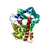
| ||||||||
|---|---|---|---|---|---|---|---|---|---|
| 1 |
| ||||||||
| Unit cell |
|
-Components
| #1: Protein | Mass: 32047.600 Da / Num. of mol.: 1 Source method: isolated from a genetically manipulated source Source: (gene. exp.) Firmicutes bacterium CAG:884 (bacteria)Gene: BN804_00706 / Production host: References: UniProt: R5MX27, phosphatidate cytidylyltransferase | ||||
|---|---|---|---|---|---|
| #2: Chemical | ChemComp-CTP /   Mass: 483.156 Da / Num. of mol.: 1 / Source method: obtained synthetically / Formula: C9H16N3O14P3 / Feature type: SUBJECT OF INVESTIGATION Mass: 483.156 Da / Num. of mol.: 1 / Source method: obtained synthetically / Formula: C9H16N3O14P3 / Feature type: SUBJECT OF INVESTIGATION | ||||
| #3: Chemical | ChemComp-MG /   Mass: 24.305 Da / Num. of mol.: 1 / Source method: obtained synthetically / Formula: Mg / Feature type: SUBJECT OF INVESTIGATION Mass: 24.305 Da / Num. of mol.: 1 / Source method: obtained synthetically / Formula: Mg / Feature type: SUBJECT OF INVESTIGATION | ||||
| #4: Chemical |   Mass: 79.904 Da / Num. of mol.: 2 / Source method: obtained synthetically / Formula: Br Mass: 79.904 Da / Num. of mol.: 2 / Source method: obtained synthetically / Formula: Br#5: Water | ChemComp-HOH / |  Mass: 18.015 Da / Num. of mol.: 2 / Source method: isolated from a natural source / Formula: H2O Mass: 18.015 Da / Num. of mol.: 2 / Source method: isolated from a natural source / Formula: H2OHas ligand of interest | Y | |
-Experimental details
-Experiment
| Experiment | Method: X-RAY DIFFRACTION / Number of used crystals: 1 |
|---|
- Sample preparation
| Crystal | Density Matthews: 4.82 Å3/Da / Density % sol: 74.49 % |
|---|---|
| Crystal grow | Temperature: 293 K / Method: vapor diffusion, sitting drop Details: 0.1M HEPES ph 7.0, 13-15% (w/v) PEG 6000, 10% (v/v) Glycerol |
-Data collection
| Diffraction | Mean temperature: 100 K / Serial crystal experiment: N |
|---|---|
| Diffraction source | Source: SYNCHROTRON / Site: SPring-8 / Beamline: BL32XU / Wavelength: 0.92 Å |
| Detector | Type: DECTRIS EIGER X 9M / Detector: PIXEL / Date: Nov 17, 2019 |
| Radiation | Protocol: SINGLE WAVELENGTH / Monochromatic (M) / Laue (L): M / Scattering type: x-ray |
| Radiation wavelength | Wavelength: 0.92 Å / Relative weight: 1 |
| Reflection | Resolution: 2.6→50 Å / Num. obs: 36822 / % possible obs: 99.7 % / Redundancy: 13.5 % / CC1/2: 0.999 / Net I/σ(I): 23.8 |
| Reflection shell | Resolution: 2.6→2.76 Å / Num. unique obs: 5901 / CC1/2: 0.722 |
- Processing
| Software |
| |||||||||||||||||||||||||||||||||||||||||||||||||
|---|---|---|---|---|---|---|---|---|---|---|---|---|---|---|---|---|---|---|---|---|---|---|---|---|---|---|---|---|---|---|---|---|---|---|---|---|---|---|---|---|---|---|---|---|---|---|---|---|---|---|
| Refinement | Method to determine structure: SAD / Resolution: 2.6→43.12 Å / SU ML: 0.45 / Cross valid method: FREE R-VALUE / σ(F): 1.36 / Phase error: 28.25 / Stereochemistry target values: ML
| |||||||||||||||||||||||||||||||||||||||||||||||||
| Solvent computation | Shrinkage radii: 0.9 Å / VDW probe radii: 1.11 Å / Solvent model: FLAT BULK SOLVENT MODEL | |||||||||||||||||||||||||||||||||||||||||||||||||
| Refinement step | Cycle: LAST / Resolution: 2.6→43.12 Å
| |||||||||||||||||||||||||||||||||||||||||||||||||
| Refine LS restraints |
| |||||||||||||||||||||||||||||||||||||||||||||||||
| LS refinement shell |
|
