 Movie
Movie Controller

[English] 日本語
 Yorodumi
- PDB-7ead: Crystal structure of beta-sheet cytochrome c prime from Thermus t... -
+ Open data

- Basic information
| Entry | Database: PDB / ID: 7ead | ||||||||||||
|---|---|---|---|---|---|---|---|---|---|---|---|---|---|
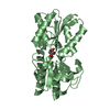
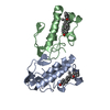
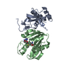
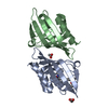
| Title | Crystal structure of beta-sheet cytochrome c prime from Thermus thermophilus. | ||||||||||||
 Components Components | Cytochrome_P460 domain-containing protein | ||||||||||||
 Keywords Keywords | ELECTRON TRANSPORT / Cytochrome c / thermophile | ||||||||||||
| Function / homology | HEME C / :  Function and homology information Function and homology information | ||||||||||||
| Biological species |   Thermus thermophilus (bacteria) Thermus thermophilus (bacteria) | ||||||||||||
| Method |  X-RAY DIFFRACTION / SYNCHROTRON / MOLECULAR REPLACEMENT / Resolution: 1.74 Å X-RAY DIFFRACTION / SYNCHROTRON / MOLECULAR REPLACEMENT / Resolution: 1.74 Å | ||||||||||||
 Authors Authors | Yoshimi, T. / Fujii, S. / Oki, H. / Igawa, T. / Adams, R.H. / Ueda, K. / Kawahara, K. / Ohkubo, T. / Hough, A.M. / Sambongi, Y. | ||||||||||||
| Funding support |  Japan, 3items Japan, 3items
| ||||||||||||
 Citation Citation | Journal: Acta Crystallogr.,Sect.F / Year: 2022 Title: Crystal structure of thermally stable homodimeric cytochrome c'-beta from Thermus thermophilus. Authors: Yoshimi, T. / Fujii, S. / Oki, H. / Igawa, T. / Adams, H.R. / Ueda, K. / Kawahara, K. / Ohkubo, T. / Hough, M.A. / Sambongi, Y. | ||||||||||||
| History |
|
- Structure visualization
| Structure viewer | Molecule: MolmilJmol/JSmol |
|---|
- Downloads & links
-Download
| PDBx/mmCIF format | 7ead.cif.gz | 45.6 KB | Display | PDBx/mmCIF format |
|---|---|---|---|---|
| PDB format | pdb7ead.ent.gz | 29.4 KB | Display | PDB format |
| PDBx/mmJSON format | 7ead.json.gz | Tree view | PDBx/mmJSON format | |
| Others |  Other downloads Other downloads |
-Validation report
| Summary document | 7ead_validation.pdf.gz | 820.4 KB | Display | wwPDB validaton report |
|---|---|---|---|---|
| Full document | 7ead_full_validation.pdf.gz | 820.5 KB | Display | |
| Data in XML | 7ead_validation.xml.gz | 8.9 KB | Display | |
| Data in CIF | 7ead_validation.cif.gz | 11.9 KB | Display | |
| Arichive directory | https://data.pdbj.org/pub/pdb/validation_reports/ea/7eadftp://data.pdbj.org/pub/pdb/validation_reports/ea/7ead | HTTPS FTP |
-Related structure data
| Related structure data |  6hihS S: Starting model for refinement |
|---|---|
| Similar structure data |


-Links
 PDBj
PDBj


- Assembly
| Deposited unit | 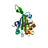
| ||||||||
|---|---|---|---|---|---|---|---|---|---|
| 1 | 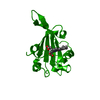
| ||||||||
| Unit cell |
| ||||||||
| Components on special symmetry positions |
|
-Components
| #1: Protein | Mass: 14942.125 Da / Num. of mol.: 1 Source method: isolated from a genetically manipulated source Source: (gene. exp.) Thermus thermophilus (bacteria) / Gene: TthHC11_22770 / Production host: |
|---|---|
| #2: Chemical | ChemComp-HEC /   Mass: 618.503 Da / Num. of mol.: 1 / Source method: obtained synthetically / Formula: C34H34FeN4O4 / Feature type: SUBJECT OF INVESTIGATION Mass: 618.503 Da / Num. of mol.: 1 / Source method: obtained synthetically / Formula: C34H34FeN4O4 / Feature type: SUBJECT OF INVESTIGATION |
| #3: Water | ChemComp-HOH /  Mass: 18.015 Da / Num. of mol.: 126 / Source method: isolated from a natural source / Formula: H2O Mass: 18.015 Da / Num. of mol.: 126 / Source method: isolated from a natural source / Formula: H2O |
| Has ligand of interest | Y |
| Has protein modification | Y |
-Experimental details
-Experiment
| Experiment | Method: X-RAY DIFFRACTION / Number of used crystals: 1 |
|---|
- Sample preparation
| Crystal | Density Matthews: 2.17 Å3/Da / Density % sol: 43.36 % |
|---|---|
| Crystal grow | Temperature: 277 K / Method: vapor diffusion, hanging drop Details: 0.1 M 4-(2-hydroxyethyl)-1-piperazineethanesulfonic acid buffer (pH 7.5) and 20% (w/v) polyethylene glycol 10,000 |
-Data collection
| Diffraction | Mean temperature: 100 K / Serial crystal experiment: N | ||||||||||||||||||||||||
|---|---|---|---|---|---|---|---|---|---|---|---|---|---|---|---|---|---|---|---|---|---|---|---|---|---|
| Diffraction source | Source: SYNCHROTRON / Site: SPring-8 / Beamline: BL26B1 / Wavelength: 1 Å | ||||||||||||||||||||||||
| Detector | Type: DECTRIS EIGER R 4M / Detector: PIXEL / Date: Jul 17, 2019 | ||||||||||||||||||||||||
| Radiation | Protocol: SINGLE WAVELENGTH / Monochromatic (M) / Laue (L): M / Scattering type: x-ray | ||||||||||||||||||||||||
| Radiation wavelength | Wavelength: 1 Å / Relative weight: 1 | ||||||||||||||||||||||||
| Reflection | Resolution: 1.74→41.15 Å / Num. obs: 13755 / % possible obs: 100 % / Redundancy: 13 % / Biso Wilson estimate: 16.98 Å2 / CC1/2: 0.998 / Rmerge(I) obs: 0.197 / Net I/σ(I): 11.3 / Num. measured all: 178741 / Scaling rejects: 23 | ||||||||||||||||||||||||
| Reflection shell | Diffraction-ID: 1
|
- Processing
| Software |
| |||||||||||||||||||||||||||||||||||||||||||||||||||||||||||||||||||||||||||||
|---|---|---|---|---|---|---|---|---|---|---|---|---|---|---|---|---|---|---|---|---|---|---|---|---|---|---|---|---|---|---|---|---|---|---|---|---|---|---|---|---|---|---|---|---|---|---|---|---|---|---|---|---|---|---|---|---|---|---|---|---|---|---|---|---|---|---|---|---|---|---|---|---|---|---|---|---|---|---|
| Refinement | Method to determine structure: MOLECULAR REPLACEMENT Starting model: 6HIH Resolution: 1.74→36.42 Å / SU ML: 0.22 / Cross valid method: THROUGHOUT / σ(F): 1.35 / Phase error: 25.88 / Stereochemistry target values: ML
| |||||||||||||||||||||||||||||||||||||||||||||||||||||||||||||||||||||||||||||
| Solvent computation | Shrinkage radii: 0.9 Å / VDW probe radii: 1.11 Å / Solvent model: FLAT BULK SOLVENT MODEL | |||||||||||||||||||||||||||||||||||||||||||||||||||||||||||||||||||||||||||||
| Displacement parameters | Biso max: 54.19 Å2 / Biso mean: 22.4242 Å2 / Biso min: 12.24 Å2 | |||||||||||||||||||||||||||||||||||||||||||||||||||||||||||||||||||||||||||||
| Refinement step | Cycle: final / Resolution: 1.74→36.42 Å
| |||||||||||||||||||||||||||||||||||||||||||||||||||||||||||||||||||||||||||||
| LS refinement shell | Refine-ID: X-RAY DIFFRACTION / Rfactor Rfree error: 0 / Total num. of bins used: 10
|
