 Movie
Movie Controller


+ Open data

- Basic information
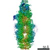
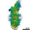
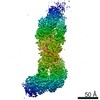
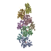
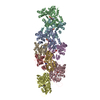
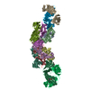
| Entry | Database: PDB / ID: 7e7i | |||||||||||||||||||||||||||
|---|---|---|---|---|---|---|---|---|---|---|---|---|---|---|---|---|---|---|---|---|---|---|---|---|---|---|---|---|
| Title | Cryo-EM structure of human ABCA4 in the apo state | |||||||||||||||||||||||||||
 Components Components | Retinal-specific phospholipid-transporting ATPase ABCA4 | |||||||||||||||||||||||||||
 Keywords Keywords | TRANSLOCASE / lipid transport / MEMBRANE PROTEIN | |||||||||||||||||||||||||||
| Function / homology |  Function and homology information Function and homology informationrod photoreceptor disc membrane / flippase activity / N-retinylidene-phosphatidylethanolamine flippase activity / Defective visual phototransduction due to ABCA4 loss of function / all-trans retinal binding / retinol transmembrane transporter activity / phospholipid transfer to membrane / : / 11-cis retinal binding / ATPase-coupled intramembrane lipid carrier activity ...rod photoreceptor disc membrane / flippase activity / N-retinylidene-phosphatidylethanolamine flippase activity / Defective visual phototransduction due to ABCA4 loss of function / all-trans retinal binding / retinol transmembrane transporter activity / phospholipid transfer to membrane / : / 11-cis retinal binding / ATPase-coupled intramembrane lipid carrier activity / retinal metabolic process / phosphatidylethanolamine flippase activity / retinoid binding / P-type phospholipid transporter / photoreceptor cell maintenance / phospholipid translocation / lipid transport / The canonical retinoid cycle in rods (twilight vision) / ATPase-coupled transmembrane transporter activity / phototransduction, visible light / photoreceptor outer segment / retinoid metabolic process / ABC-type transporter activity / visual perception / transmembrane transport / ABC-family protein mediated transport / photoreceptor disc membrane / cytoplasmic vesicle / GTPase activity / endoplasmic reticulum / ATP hydrolysis activity / ATP binding / membrane / plasma membrane Similarity search - Function | |||||||||||||||||||||||||||
| Biological species |  Homo sapiens (human) Homo sapiens (human) | |||||||||||||||||||||||||||
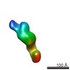
| Method | ELECTRON MICROSCOPY / single particle reconstruction / cryo EM / Resolution: 3.3 Å | |||||||||||||||||||||||||||
 Authors Authors | Xie, T. / Zhang, Z.K. / Gong, X. | |||||||||||||||||||||||||||
 Citation Citation | Journal: Nat Commun / Year: 2021 Title: Structural basis of substrate recognition and translocation by human ABCA4. Authors: Tian Xie / Zike Zhang / Qi Fang / Bowen Du / Xin Gong /  Abstract: Human ATP-binding cassette (ABC) subfamily A (ABCA) transporters mediate the transport of various lipid compounds across the membrane. Mutations in human ABCA transporters have been described to ...Human ATP-binding cassette (ABC) subfamily A (ABCA) transporters mediate the transport of various lipid compounds across the membrane. Mutations in human ABCA transporters have been described to cause severe hereditary disorders associated with impaired lipid transport. However, little is known about the mechanistic details of substrate recognition and translocation by ABCA transporters. Here, we present three cryo-EM structures of human ABCA4, a retina-specific ABCA transporter, in distinct functional states at resolutions of 3.3-3.4 Å. In the nucleotide-free state, the two transmembrane domains (TMDs) exhibit a lateral-opening conformation, allowing the lateral entry of substrate from the lipid bilayer. The N-retinylidene-phosphatidylethanolamine (NRPE), the physiological lipid substrate of ABCA4, is sandwiched between the two TMDs in the luminal leaflet and is further stabilized by an extended loop from extracellular domain 1. In the ATP-bound state, the two TMDs display a closed conformation, which precludes the substrate binding. Our study provides a molecular basis to understand the mechanism of ABCA4-mediated NRPE recognition and translocation, and suggests a common 'lateral access and extrusion' mechanism for ABCA-mediated lipid transport. | |||||||||||||||||||||||||||
| History |
|
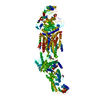
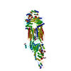
- Structure visualization
| Movie |
Movie viewer |
|---|---|
| Structure viewer | Molecule: MolmilJmol/JSmol |
- Downloads & links
-Download
| PDBx/mmCIF format | 7e7i.cif.gz | 378.9 KB | Display | PDBx/mmCIF format |
|---|---|---|---|---|
| PDB format | pdb7e7i.ent.gz | 291.8 KB | Display | PDB format |
| PDBx/mmJSON format | 7e7i.json.gz | Tree view | PDBx/mmJSON format | |
| Others |  Other downloads Other downloads |
-Validation report
| Arichive directory | https://data.pdbj.org/pub/pdb/validation_reports/e7/7e7iftp://data.pdbj.org/pub/pdb/validation_reports/e7/7e7i | HTTPS FTP |
|---|
-Related structure data
| Related structure data | 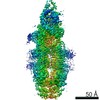 31000MC 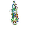 7e7oC 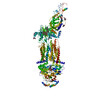 7e7qC M: map data used to model this data C: citing same article ( |
|---|---|
| Similar structure data |
-Links
 PDBj
PDBj


- Assembly
| Deposited unit | 
|
|---|---|
| 1 |
|
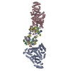
-Components
| #1: Protein | Mass: 261143.547 Da / Num. of mol.: 1 Source method: isolated from a genetically manipulated source Source: (gene. exp.) Homo sapiens (human) / Gene: ABCA4, ABCR / Production host: Homo sapiens (human)References: UniProt: P78363, P-type phospholipid transporter | ||||||||
|---|---|---|---|---|---|---|---|---|---|
| #2: Polysaccharide | Source method: isolated from a genetically manipulated source #3: Sugar | ChemComp-NAG /   Type: D-saccharide, beta linking / Mass: 221.208 Da / Num. of mol.: 7 Type: D-saccharide, beta linking / Mass: 221.208 Da / Num. of mol.: 7Source method: isolated from a genetically manipulated source Formula: C8H15NO6 #4: Chemical |   Mass: 748.065 Da / Num. of mol.: 2 Mass: 748.065 Da / Num. of mol.: 2Source method: isolated from a genetically manipulated source Formula: C41H82NO8P / Feature type: SUBJECT OF INVESTIGATION / Comment: phospholipid*YM Has ligand of interest | Y | Has protein modification | Y | |
-Experimental details
-Experiment
| Experiment | Method: ELECTRON MICROSCOPY |
|---|---|
| EM experiment | Aggregation state: PARTICLE / 3D reconstruction method: single particle reconstruction |
- Sample preparation
| Component | Name: ABCA4 / Type: COMPLEX / Entity ID: #1 / Source: RECOMBINANT |
|---|---|
| Source (natural) | Organism: Homo sapiens (human) |
| Source (recombinant) | Organism: Homo sapiens (human) |
| Buffer solution | pH: 7.5 |
| Specimen | Embedding applied: NO / Shadowing applied: NO / Staining applied: NO / Vitrification applied: YES |
| Vitrification | Cryogen name: ETHANE |
- Electron microscopy imaging
| Experimental equipment |  Model: Titan Krios / Image courtesy: FEI Company |
|---|---|
| Microscopy | Model: FEI TITAN KRIOS |
| Electron gun | Electron source:  FIELD EMISSION GUN / Accelerating voltage: 300 kV / Illumination mode: FLOOD BEAM FIELD EMISSION GUN / Accelerating voltage: 300 kV / Illumination mode: FLOOD BEAM |
| Electron lens | Mode: BRIGHT FIELD |
| Image recording | Electron dose: 50 e/Å2 / Film or detector model: GATAN K2 SUMMIT (4k x 4k) |
- Processing
| Software | Name: PHENIX / Version: 1.18.1_3865: / Classification: refinement | ||||||||||||||||||||||||
|---|---|---|---|---|---|---|---|---|---|---|---|---|---|---|---|---|---|---|---|---|---|---|---|---|---|
| EM software | Name: PHENIX / Category: model refinement | ||||||||||||||||||||||||
| CTF correction | Type: NONE | ||||||||||||||||||||||||
| 3D reconstruction | Resolution: 3.3 Å / Resolution method: FSC 0.143 CUT-OFF / Num. of particles: 205597 / Symmetry type: POINT | ||||||||||||||||||||||||
| Refine LS restraints |
|
