 Movie
Movie Controller

[English] 日本語
 Yorodumi
- PDB-7e65: The crystal structure of peptidoglycan peptidase in complex with ... -
+ Open data

- Basic information
| Entry | Database: PDB / ID: 7.0E+65 | ||||||
|---|---|---|---|---|---|---|---|
| Title | The crystal structure of peptidoglycan peptidase in complex with inhibitor 3 | ||||||
 Components Components | Peptidase M23 | ||||||
 Keywords Keywords | HYDROLASE/INHIBITOR / HYDROLASE-INHIBITOR COMPLEX | ||||||
| Function / homology | Glucose Permease (Domain IIA) / Glucose Permease (Domain IIA) / Peptidase M23 / Peptidase family M23 / Duplicated hybrid motif / Distorted Sandwich / Mainly Beta / Chem-HXO / Peptidase M23 Function and homology information Function and homology information | ||||||
| Biological species |   Campylobacter jejuni (Campylobacter) Campylobacter jejuni (Campylobacter) | ||||||
| Method |  X-RAY DIFFRACTION / SYNCHROTRON / MOLECULAR REPLACEMENT / Resolution: 2.65 Å X-RAY DIFFRACTION / SYNCHROTRON / MOLECULAR REPLACEMENT / Resolution: 2.65 Å | ||||||
 Authors Authors | Choi, Y. / Min, K.J. / Yoon, H.J. / Lee, H.H. | ||||||
 Citation Citation | Journal: Commun Biol / Year: 2022 Title: Structure-based inhibitor design for reshaping bacterial morphology Authors: Choi, Y. / Park, J.S. / Kim, J. / Min, K. / Mahasenan, K. / Kim, C. / Yoon, H.J. / Lim, S. / Cheon, D.H. / Lee, Y. / Ryu, S. / Mobashery, S. / Kim, B.M. / Lee, H.H. | ||||||
| History |
|
- Structure visualization
| Structure viewer | Molecule: MolmilJmol/JSmol |
|---|
- Downloads & links
-Download
| PDBx/mmCIF format | 7e65.cif.gz | 116.3 KB | Display | PDBx/mmCIF format |
|---|---|---|---|---|
| PDB format | pdb7e65.ent.gz | 87.4 KB | Display | PDB format |
| PDBx/mmJSON format | 7e65.json.gz | Tree view | PDBx/mmJSON format | |
| Others |  Other downloads Other downloads |
-Validation report
| Summary document | 7e65_validation.pdf.gz | 1.2 MB | Display | wwPDB validaton report |
|---|---|---|---|---|
| Full document | 7e65_full_validation.pdf.gz | 1.3 MB | Display | |
| Data in XML | 7e65_validation.xml.gz | 21.2 KB | Display | |
| Data in CIF | 7e65_validation.cif.gz | 29 KB | Display | |
| Arichive directory | https://data.pdbj.org/pub/pdb/validation_reports/e6/7e65ftp://data.pdbj.org/pub/pdb/validation_reports/e6/7e65 | HTTPS FTP |
-Related structure data
| Related structure data |  7e60C 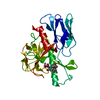 7e61C 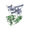 7e63C 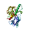 7e64C 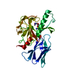 7e66C 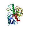 7e67C 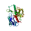 7e69C  6jmzS C: citing same article ( S: Starting model for refinement |
|---|---|
| Similar structure data |


-Links
 PDBj
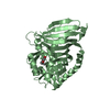
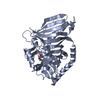
PDBj- Assembly
| Deposited unit | 
| ||||||||||||||||||
|---|---|---|---|---|---|---|---|---|---|---|---|---|---|---|---|---|---|---|---|
| 1 | 
| ||||||||||||||||||
| 2 | 
| ||||||||||||||||||
| Unit cell |
| ||||||||||||||||||
| Noncrystallographic symmetry (NCS) | NCS domain:
NCS domain segments: Component-ID: _ / Ens-ID: 1 / Beg auth comp-ID: MET / Beg label comp-ID: MET / End auth comp-ID: LEU / End label comp-ID: LEU / Refine code: _ / Auth seq-ID: 20 - 274 / Label seq-ID: 1 - 255
|
-Components
| #1: Protein | Mass: 28463.752 Da / Num. of mol.: 2 / Mutation: H247A Source method: isolated from a genetically manipulated source Source: (gene. exp.) Campylobacter jejuni (Campylobacter) / Gene: A8118_01115 / Production host: #2: Chemical |   Mass: 65.409 Da / Num. of mol.: 2 / Source method: obtained synthetically / Formula: Zn / Feature type: SUBJECT OF INVESTIGATION Mass: 65.409 Da / Num. of mol.: 2 / Source method: obtained synthetically / Formula: Zn / Feature type: SUBJECT OF INVESTIGATION#3: Chemical | ChemComp-HXO / ( | 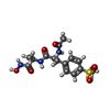  Mass: 372.397 Da / Num. of mol.: 1 / Source method: obtained synthetically / Formula: C14H20N4O6S / Feature type: SUBJECT OF INVESTIGATION Mass: 372.397 Da / Num. of mol.: 1 / Source method: obtained synthetically / Formula: C14H20N4O6S / Feature type: SUBJECT OF INVESTIGATION#4: Water | ChemComp-HOH / |  Mass: 18.015 Da / Num. of mol.: 91 / Source method: isolated from a natural source / Formula: H2O Mass: 18.015 Da / Num. of mol.: 91 / Source method: isolated from a natural source / Formula: H2OHas ligand of interest | Y | |
|---|
-Experimental details
-Experiment
| Experiment | Method: X-RAY DIFFRACTION / Number of used crystals: 1 |
|---|
- Sample preparation
| Crystal | Density Matthews: 2.28 Å3/Da / Density % sol: 45.96 % |
|---|---|
| Crystal grow | Temperature: 296 K / Method: vapor diffusion, sitting drop Details: 0.1 M sodium citrate tribasic dihydrate pH 5.5 and 22% PEG 1,000 |
-Data collection
| Diffraction | Mean temperature: 100 K / Serial crystal experiment: N | |||||||||||||||||||||||||||||||||||||||||||||||||||||||||||||||||||||||||||||||||||||||||||||||||||||||||||||||||||||||||||||||||||||||||||||||||||||||||||||||||||||||||||||||||||||||||||||
|---|---|---|---|---|---|---|---|---|---|---|---|---|---|---|---|---|---|---|---|---|---|---|---|---|---|---|---|---|---|---|---|---|---|---|---|---|---|---|---|---|---|---|---|---|---|---|---|---|---|---|---|---|---|---|---|---|---|---|---|---|---|---|---|---|---|---|---|---|---|---|---|---|---|---|---|---|---|---|---|---|---|---|---|---|---|---|---|---|---|---|---|---|---|---|---|---|---|---|---|---|---|---|---|---|---|---|---|---|---|---|---|---|---|---|---|---|---|---|---|---|---|---|---|---|---|---|---|---|---|---|---|---|---|---|---|---|---|---|---|---|---|---|---|---|---|---|---|---|---|---|---|---|---|---|---|---|---|---|---|---|---|---|---|---|---|---|---|---|---|---|---|---|---|---|---|---|---|---|---|---|---|---|---|---|---|---|---|---|---|---|
| Diffraction source | Source: SYNCHROTRON / Site: PAL/PLS  / Beamline: 5C (4A) / Wavelength: 0.9796 Å / Beamline: 5C (4A) / Wavelength: 0.9796 Å | |||||||||||||||||||||||||||||||||||||||||||||||||||||||||||||||||||||||||||||||||||||||||||||||||||||||||||||||||||||||||||||||||||||||||||||||||||||||||||||||||||||||||||||||||||||||||||||
| Detector | Type: DECTRIS EIGER X 9M / Detector: PIXEL / Date: Dec 14, 2020 | |||||||||||||||||||||||||||||||||||||||||||||||||||||||||||||||||||||||||||||||||||||||||||||||||||||||||||||||||||||||||||||||||||||||||||||||||||||||||||||||||||||||||||||||||||||||||||||
| Radiation | Protocol: SINGLE WAVELENGTH / Monochromatic (M) / Laue (L): M / Scattering type: x-ray | |||||||||||||||||||||||||||||||||||||||||||||||||||||||||||||||||||||||||||||||||||||||||||||||||||||||||||||||||||||||||||||||||||||||||||||||||||||||||||||||||||||||||||||||||||||||||||||
| Radiation wavelength | Wavelength: 0.9796 Å / Relative weight: 1 | |||||||||||||||||||||||||||||||||||||||||||||||||||||||||||||||||||||||||||||||||||||||||||||||||||||||||||||||||||||||||||||||||||||||||||||||||||||||||||||||||||||||||||||||||||||||||||||
| Reflection | Resolution: 2.65→70 Å / Num. obs: 15653 / % possible obs: 99.8 % / Redundancy: 12.3 % / Rmerge(I) obs: 0.184 / Rpim(I) all: 0.054 / Rrim(I) all: 0.192 / Χ2: 0.961 / Net I/σ(I): 5.1 / Num. measured all: 191812 | |||||||||||||||||||||||||||||||||||||||||||||||||||||||||||||||||||||||||||||||||||||||||||||||||||||||||||||||||||||||||||||||||||||||||||||||||||||||||||||||||||||||||||||||||||||||||||||
| Reflection shell | Diffraction-ID: 1
|
- Processing
| Software |
| ||||||||||||||||||||||||||||||||||||||||||||||||||||||||||||
|---|---|---|---|---|---|---|---|---|---|---|---|---|---|---|---|---|---|---|---|---|---|---|---|---|---|---|---|---|---|---|---|---|---|---|---|---|---|---|---|---|---|---|---|---|---|---|---|---|---|---|---|---|---|---|---|---|---|---|---|---|---|
| Refinement | Method to determine structure: MOLECULAR REPLACEMENT Starting model: 6JMZ Resolution: 2.65→67.58 Å / Cor.coef. Fo:Fc: 0.94 / Cor.coef. Fo:Fc free: 0.88 / SU B: 14.331 / SU ML: 0.306 / Cross valid method: THROUGHOUT / σ(F): 0 / ESU R Free: 0.418 / Stereochemistry target values: MAXIMUM LIKELIHOOD Details: HYDROGENS HAVE BEEN ADDED IN THE RIDING POSITIONS U VALUES : REFINED INDIVIDUALLY
| ||||||||||||||||||||||||||||||||||||||||||||||||||||||||||||
| Solvent computation | Ion probe radii: 0.8 Å / Shrinkage radii: 0.8 Å / VDW probe radii: 1.2 Å / Solvent model: MASK | ||||||||||||||||||||||||||||||||||||||||||||||||||||||||||||
| Displacement parameters | Biso max: 154.98 Å2 / Biso mean: 45.936 Å2 / Biso min: 12.57 Å2
| ||||||||||||||||||||||||||||||||||||||||||||||||||||||||||||
| Refinement step | Cycle: final / Resolution: 2.65→67.58 Å
| ||||||||||||||||||||||||||||||||||||||||||||||||||||||||||||
| Refine LS restraints |
| ||||||||||||||||||||||||||||||||||||||||||||||||||||||||||||
| Refine LS restraints NCS | Ens-ID: 1 / Number: 14816 / Refine-ID: X-RAY DIFFRACTION / Type: interatomic distance / Rms dev position: 0.12 Å / Weight position: 0.05
| ||||||||||||||||||||||||||||||||||||||||||||||||||||||||||||
| LS refinement shell | Resolution: 2.65→2.718 Å / Rfactor Rfree error: 0
|
