 Movie
Movie Controller

[English] 日本語
 Yorodumi
- PDB-7e3u: Crystal structure of the Pseudomonas aeruginosa dihydropyrimidina... -
+ Open data

- Basic information
| Entry | Database: PDB / ID: 7e3u | ||||||
|---|---|---|---|---|---|---|---|
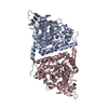
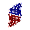
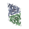
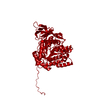
| Title | Crystal structure of the Pseudomonas aeruginosa dihydropyrimidinase complexed with 5-AU | ||||||
 Components Components | D-hydantoinase/dihydropyrimidinase | ||||||
 Keywords Keywords | HYDROLASE / dihydropyrimidinase | ||||||
| Function / homology |  Function and homology information Function and homology informationdihydropyrimidinase / pyrimidine-containing compound metabolic process / dihydropyrimidinase activity / hydrolase activity, acting on carbon-nitrogen (but not peptide) bonds, in cyclic amides / nucleobase-containing small molecule metabolic process / metal ion binding / cytosol Similarity search - Function | ||||||
| Biological species |   Pseudomonas aeruginosa (bacteria) Pseudomonas aeruginosa (bacteria) | ||||||
| Method |  X-RAY DIFFRACTION / SYNCHROTRON / MOLECULAR REPLACEMENT / Resolution: 2.159 Å X-RAY DIFFRACTION / SYNCHROTRON / MOLECULAR REPLACEMENT / Resolution: 2.159 Å | ||||||
 Authors Authors | Yang, Y.C. / Luo, R.H. / Huang, Y.H. / Huang, C.Y. / Lin, E.S. | ||||||
 Citation Citation | Journal: Bioinorg Chem Appl / Year: 2022 Title: Molecular Insights into How the Dimetal Center in Dihydropyrimidinase Can Bind the Thymine Antagonist 5-Aminouracil: A Different Binding Mode from the Anticancer Drug 5-Fluorouracil. Authors: Lin, E.S. / Luo, R.H. / Yang, Y.C. / Huang, C.Y. | ||||||
| History |
|
- Structure visualization
| Structure viewer | Molecule: MolmilJmol/JSmol |
|---|
- Downloads & links
-Download
| PDBx/mmCIF format | 7e3u.cif.gz | 204.1 KB | Display | PDBx/mmCIF format |
|---|---|---|---|---|
| PDB format | pdb7e3u.ent.gz | 160.5 KB | Display | PDB format |
| PDBx/mmJSON format | 7e3u.json.gz | Tree view | PDBx/mmJSON format | |
| Others |  Other downloads Other downloads |
-Validation report
| Arichive directory | https://data.pdbj.org/pub/pdb/validation_reports/e3/7e3uftp://data.pdbj.org/pub/pdb/validation_reports/e3/7e3u | HTTPS FTP |
|---|
-Related structure data
| Related structure data |  5e5cS S: Starting model for refinement |
|---|---|
| Similar structure data |


-Links
 PDBj
PDBj

- Assembly
| Deposited unit | 
| ||||||||||||
|---|---|---|---|---|---|---|---|---|---|---|---|---|---|
| 1 |
| ||||||||||||
| Unit cell |
| ||||||||||||
| Components on special symmetry positions |
|
-Components
| #1: Protein | Mass: 52315.844 Da / Num. of mol.: 2 Source method: isolated from a genetically manipulated source Source: (gene. exp.) Pseudomonas aeruginosa (strain ATCC 15692 / DSM 22644 / CIP 104116 / JCM 14847 / LMG 12228 / 1C / PRS 101 / PAO1) (bacteria)Strain: ATCC 15692 / DSM 22644 / CIP 104116 / JCM 14847 / LMG 12228 / 1C / PRS 101 / PAO1 Gene: dht, PA0441 Production host: References: UniProt: Q9I676, dihydropyrimidinase #2: Chemical | ChemComp-WBU / |   Mass: 127.101 Da / Num. of mol.: 1 / Source method: obtained synthetically / Formula: C4H5N3O2 / Feature type: SUBJECT OF INVESTIGATION Mass: 127.101 Da / Num. of mol.: 1 / Source method: obtained synthetically / Formula: C4H5N3O2 / Feature type: SUBJECT OF INVESTIGATION#3: Chemical | ChemComp-ZN /   Mass: 65.409 Da / Num. of mol.: 4 / Source method: obtained synthetically / Formula: Zn / Feature type: SUBJECT OF INVESTIGATION Mass: 65.409 Da / Num. of mol.: 4 / Source method: obtained synthetically / Formula: Zn / Feature type: SUBJECT OF INVESTIGATION#4: Water | ChemComp-HOH / |  Mass: 18.015 Da / Num. of mol.: 459 / Source method: isolated from a natural source / Formula: H2O Mass: 18.015 Da / Num. of mol.: 459 / Source method: isolated from a natural source / Formula: H2OHas ligand of interest | Y | |
|---|
-Experimental details
-Experiment
| Experiment | Method: X-RAY DIFFRACTION / Number of used crystals: 1 |
|---|
- Sample preparation
| Crystal | Density Matthews: 2.83 Å3/Da / Density % sol: 56.49 % |
|---|---|
| Crystal grow | Temperature: 298 K / Method: vapor diffusion, hanging drop Details: 16%PEG8000, 100mM HEPES pH7.5, 200mM Calcium Acetate |
-Data collection
| Diffraction | Mean temperature: 298 K / Serial crystal experiment: N |
|---|---|
| Diffraction source | Source: SYNCHROTRON / Site: NSRRC  / Beamline: BL13B1 / Wavelength: 1 Å / Beamline: BL13B1 / Wavelength: 1 Å |
| Detector | Type: ADSC QUANTUM 210r / Detector: CCD / Date: Dec 17, 2020 |
| Radiation | Protocol: SINGLE WAVELENGTH / Monochromatic (M) / Laue (L): M / Scattering type: x-ray |
| Radiation wavelength | Wavelength: 1 Å / Relative weight: 1 |
| Reflection | Resolution: 2.159→30 Å / Num. obs: 64183 / % possible obs: 99.9 % / Redundancy: 4.9 % / Rmerge(I) obs: 0.066 / Net I/σ(I): 3.1 |
| Reflection shell | Resolution: 2.159→2.24 Å / Redundancy: 4.9 % / Mean I/σ(I) obs: 23.7 / Num. unique obs: 6340 / CC1/2: 0.866 / % possible all: 99.9 |
- Processing
| Software |
| ||||||||||||||||||||||||||||||||||||||||||||||||||||||||||||||||||||||||||||||||||||||||||||||||||||||||||||||||||||||||||||||||||||||||||||||||
|---|---|---|---|---|---|---|---|---|---|---|---|---|---|---|---|---|---|---|---|---|---|---|---|---|---|---|---|---|---|---|---|---|---|---|---|---|---|---|---|---|---|---|---|---|---|---|---|---|---|---|---|---|---|---|---|---|---|---|---|---|---|---|---|---|---|---|---|---|---|---|---|---|---|---|---|---|---|---|---|---|---|---|---|---|---|---|---|---|---|---|---|---|---|---|---|---|---|---|---|---|---|---|---|---|---|---|---|---|---|---|---|---|---|---|---|---|---|---|---|---|---|---|---|---|---|---|---|---|---|---|---|---|---|---|---|---|---|---|---|---|---|---|---|---|---|
| Refinement | Method to determine structure: MOLECULAR REPLACEMENT Starting model: 5E5C Resolution: 2.159→28.012 Å / SU ML: 0.26 / Cross valid method: THROUGHOUT / σ(F): 1.35 / Phase error: 23.46 / Stereochemistry target values: ML
| ||||||||||||||||||||||||||||||||||||||||||||||||||||||||||||||||||||||||||||||||||||||||||||||||||||||||||||||||||||||||||||||||||||||||||||||||
| Solvent computation | Shrinkage radii: 0.9 Å / VDW probe radii: 1.11 Å / Solvent model: FLAT BULK SOLVENT MODEL | ||||||||||||||||||||||||||||||||||||||||||||||||||||||||||||||||||||||||||||||||||||||||||||||||||||||||||||||||||||||||||||||||||||||||||||||||
| Displacement parameters | Biso max: 81.35 Å2 / Biso mean: 36.4665 Å2 / Biso min: 14.4 Å2 | ||||||||||||||||||||||||||||||||||||||||||||||||||||||||||||||||||||||||||||||||||||||||||||||||||||||||||||||||||||||||||||||||||||||||||||||||
| Refinement step | Cycle: final / Resolution: 2.159→28.012 Å
| ||||||||||||||||||||||||||||||||||||||||||||||||||||||||||||||||||||||||||||||||||||||||||||||||||||||||||||||||||||||||||||||||||||||||||||||||
| LS refinement shell | Refine-ID: X-RAY DIFFRACTION / Rfactor Rfree error: 0
|
