 Movie
Movie Controller

[English] 日本語
 Yorodumi
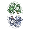
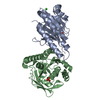
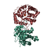
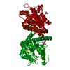
- PDB-7e31: Crystal structure of a novel alpha/beta hydrolase mutant in apo form -
+ Open data

- Basic information
| Entry | Database: PDB / ID: 7.0E+31 | ||||||
|---|---|---|---|---|---|---|---|
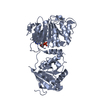
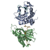
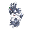
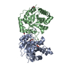
| Title | Crystal structure of a novel alpha/beta hydrolase mutant in apo form | ||||||
 Components Components | alpha/beta hydrolase | ||||||
 Keywords Keywords | HYDROLASE / alpha/beta dehydrogenase | ||||||
| Function / homology | TRIETHYLENE GLYCOL Function and homology information Function and homology information | ||||||
| Biological species | unidentified (others) | ||||||
| Method |  X-RAY DIFFRACTION / SYNCHROTRON / MOLECULAR REPLACEMENT / Resolution: 1.38 Å X-RAY DIFFRACTION / SYNCHROTRON / MOLECULAR REPLACEMENT / Resolution: 1.38 Å | ||||||
 Authors Authors | Gao, J. / Han, X. / Zheng, Y.Y. / Liu, W.D. | ||||||
| Funding support |  China, 1items China, 1items
| ||||||
 Citation Citation | Journal: Acs Catalysis / Year: 2022 Title: Multiple Substrate Binding Mode-Guided Engineering of a Thermophilic PET Hydrolase. Authors: Pfaff, L. / Gao, J. / Li, Z. / Jackering, A. / Weber, G. / Mican, J. / Chen, Y. / Dong, W. / Han, X. / Feiler, C.G. / Ao, Y.F. / Badenhorst, C.P.S. / Bednar, D. / Palm, G.J. / Lammers, M. / ...Authors: Pfaff, L. / Gao, J. / Li, Z. / Jackering, A. / Weber, G. / Mican, J. / Chen, Y. / Dong, W. / Han, X. / Feiler, C.G. / Ao, Y.F. / Badenhorst, C.P.S. / Bednar, D. / Palm, G.J. / Lammers, M. / Damborsky, J. / Strodel, B. / Liu, W. / Bornscheuer, U.T. / Wei, R. | ||||||
| History |
|
- Structure visualization
| Structure viewer | Molecule: MolmilJmol/JSmol |
|---|
- Downloads & links
-Download
| PDBx/mmCIF format | 7e31.cif.gz | 127.7 KB | Display | PDBx/mmCIF format |
|---|---|---|---|---|
| PDB format | pdb7e31.ent.gz | 96 KB | Display | PDB format |
| PDBx/mmJSON format | 7e31.json.gz | Tree view | PDBx/mmJSON format | |
| Others |  Other downloads Other downloads |
-Validation report
| Arichive directory | https://data.pdbj.org/pub/pdb/validation_reports/e3/7e31ftp://data.pdbj.org/pub/pdb/validation_reports/e3/7e31 | HTTPS FTP |
|---|
-Related structure data
| Related structure data |  7cuvC 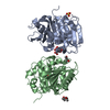 7e30C  7w66C  7w69C  7w6cC  7w6oC 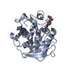 7w6qC  5zrqS S: Starting model for refinement C: citing same article ( |
|---|---|
| Similar structure data |


-Links
 PDBj
PDBj- Assembly
| Deposited unit | 
| |||||||||||||||||||||
|---|---|---|---|---|---|---|---|---|---|---|---|---|---|---|---|---|---|---|---|---|---|---|
| 1 |
| |||||||||||||||||||||
| Unit cell |
| |||||||||||||||||||||
| Noncrystallographic symmetry (NCS) | NCS domain:
NCS domain segments: Component-ID: 1 / Ens-ID: 1 / Beg auth comp-ID: GLU / Beg label comp-ID: GLU / End auth comp-ID: PHE / End label comp-ID: PHE / Auth seq-ID: 1 - 258 / Label seq-ID: 1 - 258
|
-Components
| #1: Protein | Mass: 27928.990 Da / Num. of mol.: 2 Source method: isolated from a genetically manipulated source Source: (gene. exp.) unidentified (others) / Plasmid: pET-28a / Production host:  #2: Chemical |   Mass: 150.173 Da / Num. of mol.: 2 / Source method: obtained synthetically / Formula: C6H14O4 / Feature type: SUBJECT OF INVESTIGATION Mass: 150.173 Da / Num. of mol.: 2 / Source method: obtained synthetically / Formula: C6H14O4 / Feature type: SUBJECT OF INVESTIGATION#3: Water | ChemComp-HOH / |  Mass: 18.015 Da / Num. of mol.: 773 / Source method: isolated from a natural source / Formula: H2O Mass: 18.015 Da / Num. of mol.: 773 / Source method: isolated from a natural source / Formula: H2OHas ligand of interest | Y | Has protein modification | Y | |
|---|
-Experimental details
-Experiment
| Experiment | Method: X-RAY DIFFRACTION / Number of used crystals: 1 |
|---|
- Sample preparation
| Crystal | Density Matthews: 2.48 Å3/Da / Density % sol: 50.38 % / Mosaicity: 0.83 ° |
|---|---|
| Crystal grow | Temperature: 295 K / Method: vapor diffusion, sitting drop / pH: 8 / Details: MPD, PEG1 500, NaAc |
-Data collection
| Diffraction | Mean temperature: 100 K / Serial crystal experiment: N | |||||||||||||||||||||||||||||||||||||||||||||||||||||||||||||||||||||||||||||||||||||||||||||||||||
|---|---|---|---|---|---|---|---|---|---|---|---|---|---|---|---|---|---|---|---|---|---|---|---|---|---|---|---|---|---|---|---|---|---|---|---|---|---|---|---|---|---|---|---|---|---|---|---|---|---|---|---|---|---|---|---|---|---|---|---|---|---|---|---|---|---|---|---|---|---|---|---|---|---|---|---|---|---|---|---|---|---|---|---|---|---|---|---|---|---|---|---|---|---|---|---|---|---|---|---|---|
| Diffraction source | Source: SYNCHROTRON / Site: SSRF / Beamline: BL19U1 / Wavelength: 1 Å | |||||||||||||||||||||||||||||||||||||||||||||||||||||||||||||||||||||||||||||||||||||||||||||||||||
| Detector | Type: DECTRIS PILATUS3 S 6M / Detector: PIXEL / Date: Oct 20, 2020 | |||||||||||||||||||||||||||||||||||||||||||||||||||||||||||||||||||||||||||||||||||||||||||||||||||
| Radiation | Monochromator: GRAPHITE / Protocol: SINGLE WAVELENGTH / Monochromatic (M) / Laue (L): M / Scattering type: x-ray | |||||||||||||||||||||||||||||||||||||||||||||||||||||||||||||||||||||||||||||||||||||||||||||||||||
| Radiation wavelength | Wavelength: 1 Å / Relative weight: 1 | |||||||||||||||||||||||||||||||||||||||||||||||||||||||||||||||||||||||||||||||||||||||||||||||||||
| Reflection | Resolution: 1.38→25 Å / Num. obs: 114533 / % possible obs: 100 % / Redundancy: 7.5 % / Rmerge(I) obs: 0.052 / Rpim(I) all: 0.019 / Rrim(I) all: 0.056 / Χ2: 1.584 / Net I/σ(I): 18.4 / Num. measured all: 862668 | |||||||||||||||||||||||||||||||||||||||||||||||||||||||||||||||||||||||||||||||||||||||||||||||||||
| Reflection shell | Diffraction-ID: 1
|
- Processing
| Software |
| |||||||||||||||||||||||||||||||||||||||||||||||||||||||||||||||||||||||||||||
|---|---|---|---|---|---|---|---|---|---|---|---|---|---|---|---|---|---|---|---|---|---|---|---|---|---|---|---|---|---|---|---|---|---|---|---|---|---|---|---|---|---|---|---|---|---|---|---|---|---|---|---|---|---|---|---|---|---|---|---|---|---|---|---|---|---|---|---|---|---|---|---|---|---|---|---|---|---|---|
| Refinement | Method to determine structure: MOLECULAR REPLACEMENT Starting model: 5ZRQ Resolution: 1.38→24.5 Å / SU ML: 0.11 / Cross valid method: THROUGHOUT / σ(F): 1.34 / Phase error: 16.28 / Stereochemistry target values: ML
| |||||||||||||||||||||||||||||||||||||||||||||||||||||||||||||||||||||||||||||
| Solvent computation | Shrinkage radii: 0.9 Å / VDW probe radii: 1.11 Å / Solvent model: FLAT BULK SOLVENT MODEL | |||||||||||||||||||||||||||||||||||||||||||||||||||||||||||||||||||||||||||||
| Displacement parameters | Biso max: 69.44 Å2 / Biso mean: 17.1473 Å2 / Biso min: 4.79 Å2 | |||||||||||||||||||||||||||||||||||||||||||||||||||||||||||||||||||||||||||||
| Refinement step | Cycle: final / Resolution: 1.38→24.5 Å
| |||||||||||||||||||||||||||||||||||||||||||||||||||||||||||||||||||||||||||||
| Refine LS restraints NCS |
| |||||||||||||||||||||||||||||||||||||||||||||||||||||||||||||||||||||||||||||
| LS refinement shell | Refine-ID: X-RAY DIFFRACTION / Rfactor Rfree error: 0 / Total num. of bins used: 10
|
