 Movie
Movie Controller


+ Open data

- Basic information
| Entry | Database: PDB / ID: 7e2v | ||||||
|---|---|---|---|---|---|---|---|
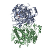
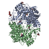
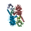
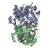
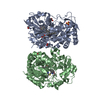
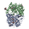
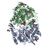
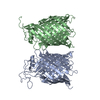
| Title | Crystal structure of MaDA-3 | ||||||
 Components Components | MaDA-3 | ||||||
 Keywords Keywords | PLANT PROTEIN / enzyme Diels-Alderase cycloaddition stereoselectivity | ||||||
| Function / homology | FLAVIN-ADENINE DINUCLEOTIDE Function and homology information Function and homology information | ||||||
| Biological species |  Morus alba (white mulberry) Morus alba (white mulberry) | ||||||
| Method |  X-RAY DIFFRACTION / SYNCHROTRON / MOLECULAR REPLACEMENT / Resolution: 2.94 Å X-RAY DIFFRACTION / SYNCHROTRON / MOLECULAR REPLACEMENT / Resolution: 2.94 Å | ||||||
 Authors Authors | Gao, L. / Du, X. / Fan, J. / Lei, X. | ||||||
| Funding support |  China, 1items China, 1items
| ||||||
 Citation Citation | Journal: Nat Catal / Year: 2021 Title: Enzymatic control of endo- and exo-stereoselective Diels-Alder reactions with broad substrate scope. Authors: Gao, L. / Zou, Y. / Liu, X. / Yang, J. / Du, X. / Wang, J. / Yu, X. / Fan, J. / Jiang, M. / Li, Y. / Houk, K.N. / Lei, X. | ||||||
| History |
|
- Structure visualization
| Structure viewer | Molecule: MolmilJmol/JSmol |
|---|
- Downloads & links
-Download
| PDBx/mmCIF format | 7e2v.cif.gz | 253.7 KB | Display | PDBx/mmCIF format |
|---|---|---|---|---|
| PDB format | pdb7e2v.ent.gz | 162.5 KB | Display | PDB format |
| PDBx/mmJSON format | 7e2v.json.gz | Tree view | PDBx/mmJSON format | |
| Others |  Other downloads Other downloads |
-Validation report
| Arichive directory | https://data.pdbj.org/pub/pdb/validation_reports/e2/7e2vftp://data.pdbj.org/pub/pdb/validation_reports/e2/7e2v | HTTPS FTP |
|---|
-Related structure data
| Related structure data |  6jqhS S: Starting model for refinement |
|---|---|
| Similar structure data |


-Links
 PDBj
PDBj- Assembly
| Deposited unit | 
| ||||||||||||||||||||||||||||||||||||||||||||||||||||||||||||||||||||||||
|---|---|---|---|---|---|---|---|---|---|---|---|---|---|---|---|---|---|---|---|---|---|---|---|---|---|---|---|---|---|---|---|---|---|---|---|---|---|---|---|---|---|---|---|---|---|---|---|---|---|---|---|---|---|---|---|---|---|---|---|---|---|---|---|---|---|---|---|---|---|---|---|---|---|
| 1 |
| ||||||||||||||||||||||||||||||||||||||||||||||||||||||||||||||||||||||||
| Unit cell |
| ||||||||||||||||||||||||||||||||||||||||||||||||||||||||||||||||||||||||
| Noncrystallographic symmetry (NCS) | NCS domain:
NCS domain segments:
NCS oper: (Code: givenMatrix: (0.445438994483, -0.838450358181, -0.313982641336), (-0.859304009601, -0.498829168544, 0.112987077548), (-0.251357755555, 0.219477692432, -0.942681717892)Vector: 24. ...NCS oper: (Code: given Matrix: (0.445438994483, -0.838450358181, -0.313982641336), Vector: |
-Components
| #1: Protein | Mass: 62387.156 Da / Num. of mol.: 2 Source method: isolated from a genetically manipulated source Source: (gene. exp.) Morus alba (white mulberry) / Production host:  Trichoplusia ni (cabbage looper) Trichoplusia ni (cabbage looper)#2: Chemical |   Mass: 785.550 Da / Num. of mol.: 2 / Source method: obtained synthetically / Formula: C27H33N9O15P2 / Comment: FAD*YM Mass: 785.550 Da / Num. of mol.: 2 / Source method: obtained synthetically / Formula: C27H33N9O15P2 / Comment: FAD*YM#3: Sugar |   Type: D-saccharide, beta linking / Mass: 221.208 Da / Num. of mol.: 2 / Source method: obtained synthetically / Formula: C8H15NO6 Type: D-saccharide, beta linking / Mass: 221.208 Da / Num. of mol.: 2 / Source method: obtained synthetically / Formula: C8H15NO6Has ligand of interest | N | Has protein modification | Y | |
|---|
-Experimental details
-Experiment
| Experiment | Method: X-RAY DIFFRACTION / Number of used crystals: 1 |
|---|
- Sample preparation
| Crystal | Density Matthews: 2.48 Å3/Da / Density % sol: 50.34 % |
|---|---|
| Crystal grow | Temperature: 291 K / Method: vapor diffusion / Details: 10% 2-propanol, 0.1 M Bicine, pH 8.5, 30% PEG 1500 |
-Data collection
| Diffraction | Mean temperature: 100 K / Serial crystal experiment: N |
|---|---|
| Diffraction source | Source: SYNCHROTRON / Site: SSRF / Beamline: BL17U / Wavelength: 0.9795 Å |
| Detector | Type: DECTRIS EIGER X 16M / Detector: PIXEL / Date: Oct 24, 2019 |
| Radiation | Protocol: SINGLE WAVELENGTH / Monochromatic (M) / Laue (L): M / Scattering type: x-ray |
| Radiation wavelength | Wavelength: 0.9795 Å / Relative weight: 1 |
| Reflection | Resolution: 2.94→43.59 Å / Num. obs: 25940 / % possible obs: 99.27 % / Redundancy: 6.5 % / Biso Wilson estimate: 53.88 Å2 / Rpim(I) all: 0.09 / Net I/σ(I): 9.9 |
| Reflection shell | Resolution: 2.95→3 Å / Num. unique obs: 1246 / CC1/2: 0.841 |
- Processing
| Software |
| ||||||||||||||||||||||||||||||||||||||||||||||||||||||||||||||||||||||
|---|---|---|---|---|---|---|---|---|---|---|---|---|---|---|---|---|---|---|---|---|---|---|---|---|---|---|---|---|---|---|---|---|---|---|---|---|---|---|---|---|---|---|---|---|---|---|---|---|---|---|---|---|---|---|---|---|---|---|---|---|---|---|---|---|---|---|---|---|---|---|---|
| Refinement | Method to determine structure: MOLECULAR REPLACEMENT Starting model: 6JQH Resolution: 2.94→43.59 Å / SU ML: 0.4997 / Cross valid method: FREE R-VALUE / σ(F): 1.35 / Phase error: 35.0523 Stereochemistry target values: GeoStd + Monomer Library + CDL v1.2
| ||||||||||||||||||||||||||||||||||||||||||||||||||||||||||||||||||||||
| Solvent computation | Shrinkage radii: 0.9 Å / VDW probe radii: 1.11 Å / Solvent model: FLAT BULK SOLVENT MODEL | ||||||||||||||||||||||||||||||||||||||||||||||||||||||||||||||||||||||
| Displacement parameters | Biso mean: 52.2 Å2 | ||||||||||||||||||||||||||||||||||||||||||||||||||||||||||||||||||||||
| Refinement step | Cycle: LAST / Resolution: 2.94→43.59 Å
| ||||||||||||||||||||||||||||||||||||||||||||||||||||||||||||||||||||||
| Refine LS restraints |
| ||||||||||||||||||||||||||||||||||||||||||||||||||||||||||||||||||||||
| Refine LS restraints NCS | Type: Torsion NCS / Rms dev position: 1.15607736955 Å | ||||||||||||||||||||||||||||||||||||||||||||||||||||||||||||||||||||||
| LS refinement shell |
|
