 Movie
Movie Controller


+ Open data

- Basic information
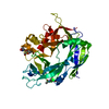
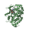
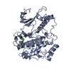
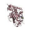
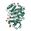
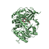
| Entry | Database: PDB / ID: 7dxm | ||||||
|---|---|---|---|---|---|---|---|
| Title | Crystal structure of DltD | ||||||
 Components Components | Protein DltD | ||||||
 Keywords Keywords | TRANSFERASE / O-acyltransferase / dlt operon | ||||||
| Function / homology | DltD / D-alanyl-lipoteichoic acid biosynthesis DltD, Firmicutes / DltD protein / lipoteichoic acid biosynthetic process / plasma membrane / Protein DltD Function and homology information Function and homology information | ||||||
| Biological species |  Streptococcus thermophilus LMG 18311 (bacteria) Streptococcus thermophilus LMG 18311 (bacteria) | ||||||
| Method |  X-RAY DIFFRACTION / SYNCHROTRON / MOLECULAR REPLACEMENT / Resolution: 2.96 Å X-RAY DIFFRACTION / SYNCHROTRON / MOLECULAR REPLACEMENT / Resolution: 2.96 Å | ||||||
 Authors Authors | Yan, X.X. / Zeng, Q. / Tian, L.F. | ||||||
 Citation Citation | Journal: Prog.Biochem.Biophys. / Year: 2022 Title: Crystal Structure of an O-acyltransfer Terminal Protein stDltD and Its Implications for dlt Operon-mediated D-alanylation of S. thermophilus. Authors: Zeng, Q. / Tian, L.F. / Liu, Y.P. / Yan, X.X. / Xu, W.Q. | ||||||
| History |
|
- Structure visualization
| Structure viewer | Molecule: MolmilJmol/JSmol |
|---|
- Downloads & links
-Download
| PDBx/mmCIF format | 7dxm.cif.gz | 316.4 KB | Display | PDBx/mmCIF format |
|---|---|---|---|---|
| PDB format | pdb7dxm.ent.gz | 259.2 KB | Display | PDB format |
| PDBx/mmJSON format | 7dxm.json.gz | Tree view | PDBx/mmJSON format | |
| Others |  Other downloads Other downloads |
-Validation report
| Arichive directory | https://data.pdbj.org/pub/pdb/validation_reports/dx/7dxmftp://data.pdbj.org/pub/pdb/validation_reports/dx/7dxm | HTTPS FTP |
|---|
-Related structure data
| Related structure data |  3bmaS S: Starting model for refinement |
|---|---|
| Similar structure data |


-Links
 PDBj
PDBj- Assembly
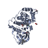
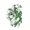
| Deposited unit | 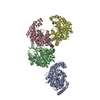
| ||||||||
|---|---|---|---|---|---|---|---|---|---|
| 1 | 
| ||||||||
| 2 | 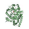
| ||||||||
| 3 | 
| ||||||||
| 4 | 
| ||||||||
| Unit cell |
|
-Components
| #1: Protein | Mass: 45829.043 Da / Num. of mol.: 4 Source method: isolated from a genetically manipulated source Source: (gene. exp.) Streptococcus thermophilus LMG 18311 (bacteria)Strain: LMG 18311 / Gene: dltD / Production host: #2: Chemical | ChemComp-SO4 /   Mass: 96.063 Da / Num. of mol.: 4 / Source method: obtained synthetically / Formula: SO4 / Feature type: SUBJECT OF INVESTIGATION Mass: 96.063 Da / Num. of mol.: 4 / Source method: obtained synthetically / Formula: SO4 / Feature type: SUBJECT OF INVESTIGATION#3: Water | ChemComp-HOH / |  Mass: 18.015 Da / Num. of mol.: 39 / Source method: isolated from a natural source / Formula: H2O Mass: 18.015 Da / Num. of mol.: 39 / Source method: isolated from a natural source / Formula: H2OHas ligand of interest | Y | |
|---|
-Experimental details
-Experiment
| Experiment | Method: X-RAY DIFFRACTION / Number of used crystals: 1 |
|---|
- Sample preparation
| Crystal | Density Matthews: 2.63 Å3/Da / Density meas: 53.22 Mg/m3 / Density % sol: 50.61 % |
|---|---|
| Crystal grow | Temperature: 293 K / Method: vapor diffusion, hanging drop / Details: 20% glycerol, 14% [w/v] PEG6000, 170 mM (NH4)2SO4) |
-Data collection
| Diffraction | Mean temperature: 81 K / Serial crystal experiment: N |
|---|---|
| Diffraction source | Source: SYNCHROTRON / Site: SSRF  / Beamline: BL19U1 / Wavelength: 0.97929 Å / Beamline: BL19U1 / Wavelength: 0.97929 Å |
| Detector | Type: DECTRIS PILATUS 6M / Detector: PIXEL / Date: Jul 20, 2020 |
| Radiation | Protocol: SINGLE WAVELENGTH / Monochromatic (M) / Laue (L): M / Scattering type: x-ray |
| Radiation wavelength | Wavelength: 0.97929 Å / Relative weight: 1 |
| Reflection | Resolution: 2.96→40 Å / Num. obs: 38815 / % possible obs: 99.2 % / Redundancy: 4 % / CC1/2: 0.984 / Rpim(I) all: 0.105 / Rrim(I) all: 0.215 / Net I/σ(I): 10.85 |
| Reflection shell | Resolution: 2.96→3.01 Å / Mean I/σ(I) obs: 1.5 / Num. unique obs: 1904 / CC1/2: 0.554 / Rpim(I) all: 0.573 |
- Processing
| Software |
| ||||||||||||||||
|---|---|---|---|---|---|---|---|---|---|---|---|---|---|---|---|---|---|
| Refinement | Method to determine structure: MOLECULAR REPLACEMENT Starting model: 3BMA Resolution: 2.96→40 Å / Cross valid method: FREE R-VALUE
| ||||||||||||||||
| Refinement step | Cycle: LAST / Resolution: 2.96→40 Å
|
