 Movie
Movie Controller

[English] 日本語
 Yorodumi
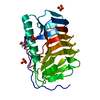
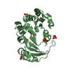
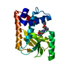
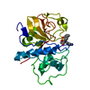
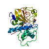
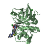
- PDB-7du4: The structure of the M.tb MazF-mt9 toxin in complex with a fragme... -
+ Open data

- Basic information
| Entry | Database: PDB / ID: 7du4 | ||||||
|---|---|---|---|---|---|---|---|
| Title | The structure of the M.tb MazF-mt9 toxin in complex with a fragment of cognate antitoxin | ||||||
 Components Components |
| ||||||
 Keywords Keywords | TOXIN / Mycobacterium tuberculosis / anti-bacterial peptide | ||||||
| Function / homology |  Function and homology information Function and homology informationsymbiont-mediated perturbation of host process / negative regulation of growth / rRNA catabolic process / mRNA catabolic process / RNA endonuclease activity / Hydrolases; Acting on ester bonds / negative regulation of translation / DNA binding Similarity search - Function | ||||||
| Biological species |  Mycobacterium tuberculosis H37Rv (bacteria) Mycobacterium tuberculosis H37Rv (bacteria) | ||||||
| Method |  X-RAY DIFFRACTION / SYNCHROTRON / MOLECULAR REPLACEMENT / Resolution: 2.18 Å X-RAY DIFFRACTION / SYNCHROTRON / MOLECULAR REPLACEMENT / Resolution: 2.18 Å | ||||||
 Authors Authors | Xie, W. / Chen, R. | ||||||
| Funding support |  China, 1items China, 1items
| ||||||
 Citation Citation | Journal: Toxins / Year: 2021 Title: Mechanistic Insight into the Peptide Binding Modes to Two M. tb MazF Toxins. Authors: Chen, R. / Zhou, J. / Xie, W. | ||||||
| History |
|
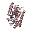
- Structure visualization
| Structure viewer | Molecule: MolmilJmol/JSmol |
|---|
- Downloads & links
-Download
| PDBx/mmCIF format | 7du4.cif.gz | 67.9 KB | Display | PDBx/mmCIF format |
|---|---|---|---|---|
| PDB format | pdb7du4.ent.gz | 40.3 KB | Display | PDB format |
| PDBx/mmJSON format | 7du4.json.gz | Tree view | PDBx/mmJSON format | |
| Others |  Other downloads Other downloads |
-Validation report
| Summary document | 7du4_validation.pdf.gz | 434 KB | Display | wwPDB validaton report |
|---|---|---|---|---|
| Full document | 7du4_full_validation.pdf.gz | 434.6 KB | Display | |
| Data in XML | 7du4_validation.xml.gz | 11 KB | Display | |
| Data in CIF | 7du4_validation.cif.gz | 14.9 KB | Display | |
| Arichive directory | https://data.pdbj.org/pub/pdb/validation_reports/du/7du4ftp://data.pdbj.org/pub/pdb/validation_reports/du/7du4 | HTTPS FTP |
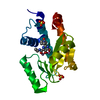
-Related structure data
| Related structure data | 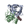 7du5C  6a6xS S: Starting model for refinement C: citing same article ( |
|---|---|
| Similar structure data |

-Links
 PDBj
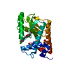
PDBj- Assembly
| Deposited unit | 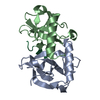
| |||||||||||||||||||||||||||||||||||||||||||||||||||
|---|---|---|---|---|---|---|---|---|---|---|---|---|---|---|---|---|---|---|---|---|---|---|---|---|---|---|---|---|---|---|---|---|---|---|---|---|---|---|---|---|---|---|---|---|---|---|---|---|---|---|---|---|
| 1 |
| |||||||||||||||||||||||||||||||||||||||||||||||||||
| Unit cell |
| |||||||||||||||||||||||||||||||||||||||||||||||||||
| Noncrystallographic symmetry (NCS) | NCS domain:
NCS domain segments: Ens-ID: 1 / End auth comp-ID: LEU / End label comp-ID: LEU
|
-Components
| #1: Protein | Mass: 14633.806 Da / Num. of mol.: 2 Source method: isolated from a genetically manipulated source Source: (gene. exp.) Mycobacterium tuberculosis H37Rv (bacteria)Strain: H37Rv / Gene: mazF7, Rv2063A / Production host: References: UniProt: P0CL62, Hydrolases; Acting on ester bonds #2: Protein/peptide | | Mass: 1635.623 Da / Num. of mol.: 1 / Source method: obtained synthetically Source: (synth.) Mycobacterium tuberculosis H37Rv (bacteria)#3: Water | ChemComp-HOH / |  Mass: 18.015 Da / Num. of mol.: 117 / Source method: isolated from a natural source / Formula: H2O Mass: 18.015 Da / Num. of mol.: 117 / Source method: isolated from a natural source / Formula: H2O |
|---|
-Experimental details
-Experiment
| Experiment | Method: X-RAY DIFFRACTION / Number of used crystals: 1 |
|---|
- Sample preparation
| Crystal | Density Matthews: 2.71 Å3/Da / Density % sol: 54.67 % |
|---|---|
| Crystal grow | Temperature: 298 K / Method: vapor diffusion, sitting drop / pH: 5 / Details: 1.5 M NaCl,0.1 M NaOAc pH 5.0 |
-Data collection
| Diffraction | Mean temperature: 100 K / Serial crystal experiment: N |
|---|---|
| Diffraction source | Source: SYNCHROTRON / Site: SSRF / Beamline: BL19U1 / Wavelength: 0.979 Å |
| Detector | Type: DECTRIS PILATUS3 S 6M / Detector: PIXEL / Date: Nov 3, 2018 |
| Radiation | Protocol: SINGLE WAVELENGTH / Monochromatic (M) / Laue (L): M / Scattering type: x-ray |
| Radiation wavelength | Wavelength: 0.979 Å / Relative weight: 1 |
| Reflection | Resolution: 2.18→50 Å / Num. obs: 17270 / % possible obs: 100 % / Redundancy: 19.2 % / Biso Wilson estimate: 27.66 Å2 / CC1/2: 0.935 / Rmerge(I) obs: 0.205 / Net I/σ(I): 13.2 |
| Reflection shell | Resolution: 2.2→2.26 Å / Rmerge(I) obs: 0.76 / Mean I/σ(I) obs: 4.15 / Num. unique obs: 1730 / CC1/2: 0.945 |
- Processing
| Software |
| |||||||||||||||||||||||||||||||||||||||||||||||||
|---|---|---|---|---|---|---|---|---|---|---|---|---|---|---|---|---|---|---|---|---|---|---|---|---|---|---|---|---|---|---|---|---|---|---|---|---|---|---|---|---|---|---|---|---|---|---|---|---|---|---|
| Refinement | Method to determine structure: MOLECULAR REPLACEMENT Starting model: 6A6X Resolution: 2.18→37.31 Å / SU ML: 0.2574 / Cross valid method: FREE R-VALUE / σ(F): 1.4 / Phase error: 22.4736 / Stereochemistry target values: CDL v1.2
| |||||||||||||||||||||||||||||||||||||||||||||||||
| Solvent computation | Shrinkage radii: 0.9 Å / VDW probe radii: 1.11 Å / Solvent model: FLAT BULK SOLVENT MODEL | |||||||||||||||||||||||||||||||||||||||||||||||||
| Displacement parameters | Biso mean: 32.49 Å2 | |||||||||||||||||||||||||||||||||||||||||||||||||
| Refinement step | Cycle: LAST / Resolution: 2.18→37.31 Å
| |||||||||||||||||||||||||||||||||||||||||||||||||
| Refine LS restraints |
| |||||||||||||||||||||||||||||||||||||||||||||||||
| LS refinement shell |
|
