 Movie
Movie Controller


+ Open data

- Basic information
| Entry | Database: PDB / ID: 7dtg | ||||||
|---|---|---|---|---|---|---|---|
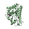
| Title | Crystal structure of lamin B1 Ig-like domain from human | ||||||
 Components Components | Lamin-B1 | ||||||
 Keywords Keywords | NUCLEAR PROTEIN / Intermediate filament / nuclear lamin B1 / Ig-like domain | ||||||
| Function / homology |  Function and homology information Function and homology informationstructural constituent of nuclear lamina / protein localization to nuclear envelope / Breakdown of the nuclear lamina / Depolymerization of the Nuclear Lamina / Nuclear Envelope Breakdown / nuclear pore localization / lamin filament / nuclear envelope organization / nuclear lamina / Initiation of Nuclear Envelope (NE) Reformation ...structural constituent of nuclear lamina / protein localization to nuclear envelope / Breakdown of the nuclear lamina / Depolymerization of the Nuclear Lamina / Nuclear Envelope Breakdown / nuclear pore localization / lamin filament / nuclear envelope organization / nuclear lamina / Initiation of Nuclear Envelope (NE) Reformation / RHOD GTPase cycle / RHOF GTPase cycle / nuclear inner membrane / Formation of Senescence-Associated Heterochromatin Foci (SAHF) / nuclear migration / Deregulated CDK5 triggers multiple neurodegenerative pathways in Alzheimer's disease models / phospholipase binding / Gene and protein expression by JAK-STAT signaling after Interleukin-12 stimulation / Meiotic synapsis / structural constituent of cytoskeleton / nuclear matrix / sequence-specific double-stranded DNA binding / nuclear envelope / heterochromatin formation / nuclear membrane / nucleoplasm / membrane / nucleus Similarity search - Function | ||||||
| Biological species |  Homo sapiens (human) Homo sapiens (human) | ||||||
| Method |  X-RAY DIFFRACTION / SYNCHROTRON / MOLECULAR REPLACEMENT / Resolution: 3.6 Å X-RAY DIFFRACTION / SYNCHROTRON / MOLECULAR REPLACEMENT / Resolution: 3.6 Å | ||||||
 Authors Authors | Ahn, J. / Lee, J. / Ha, N.-C. | ||||||
 Citation Citation | Journal: Biochem.Biophys.Res.Commun. / Year: 2021 Title: Beta-strand-mediated dimeric formation of the Ig-like domains of human lamin A/C and B1. Authors: Ahn, J. / Lee, J. / Jeong, S. / Kang, S.M. / Park, B.J. / Ha, N.C. | ||||||
| History |
|
- Structure visualization
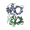
| Structure viewer | Molecule: MolmilJmol/JSmol |
|---|
- Downloads & links
-Download
| PDBx/mmCIF format | 7dtg.cif.gz | 178.4 KB | Display | PDBx/mmCIF format |
|---|---|---|---|---|
| PDB format | pdb7dtg.ent.gz | 115.1 KB | Display | PDB format |
| PDBx/mmJSON format | 7dtg.json.gz | Tree view | PDBx/mmJSON format | |
| Others |  Other downloads Other downloads |
-Validation report
| Arichive directory | https://data.pdbj.org/pub/pdb/validation_reports/dt/7dtgftp://data.pdbj.org/pub/pdb/validation_reports/dt/7dtg | HTTPS FTP |
|---|
-Related structure data
| Related structure data | 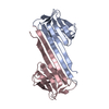 7crgC  3umnS S: Starting model for refinement C: citing same article ( |
|---|---|
| Similar structure data |


-Links
 PDBj
PDBj


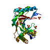
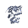
- Assembly
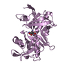
| Deposited unit | 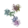
| ||||||||||||
|---|---|---|---|---|---|---|---|---|---|---|---|---|---|
| 1 | 
| ||||||||||||
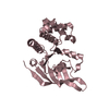
| 2 | 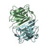
| ||||||||||||
| 3 | 
| ||||||||||||
| Unit cell |
|
-Components
| #1: Protein | Mass: 16346.178 Da / Num. of mol.: 6 / Fragment: Ig-like domain Source method: isolated from a genetically manipulated source Source: (gene. exp.) Homo sapiens (human) / Gene: LMNB1, LMN2, LMNB / Production host:  |
|---|
-Experimental details
-Experiment
| Experiment | Method: X-RAY DIFFRACTION / Number of used crystals: 1 |
|---|
- Sample preparation
| Crystal | Density Matthews: 3.94 Å3/Da / Density % sol: 68.75 % |
|---|---|
| Crystal grow | Temperature: 287.15 K / Method: vapor diffusion, hanging drop / Details: magnesium nitrate, PEG 3350 |
-Data collection
| Diffraction | Mean temperature: 80 K / Serial crystal experiment: N |
|---|---|
| Diffraction source | Source: SYNCHROTRON / Site: PAL/PLS  / Beamline: 5C (4A) / Wavelength: 0.979 Å / Beamline: 5C (4A) / Wavelength: 0.979 Å |
| Detector | Type: DECTRIS EIGER X 9M / Detector: PIXEL / Date: Aug 6, 2020 |
| Radiation | Protocol: SINGLE WAVELENGTH / Monochromatic (M) / Laue (L): M / Scattering type: x-ray |
| Radiation wavelength | Wavelength: 0.979 Å / Relative weight: 1 |
| Reflection | Resolution: 3.6→50 Å / Num. obs: 17863 / % possible obs: 97.1 % / Redundancy: 7.4 % / Biso Wilson estimate: 25.16 Å2 / CC1/2: 0.997 / CC star: 0.999 / Rpim(I) all: 0.025 / Rrim(I) all: 0.08 / Net I/σ(I): 17.95 |
| Reflection shell | Resolution: 3.6→3.66 Å / Redundancy: 4.2 % / Mean I/σ(I) obs: 3.3 / Num. unique obs: 818 / CC1/2: 0.634 / CC star: 0.881 / Rpim(I) all: 0.15 / Rrim(I) all: 0.36 / % possible all: 91.1 |
- Processing
| Software |
| |||||||||||||||||||||||||||||||||||||||||||||||||||||||||||||||||||||||||||||||||||||||||||
|---|---|---|---|---|---|---|---|---|---|---|---|---|---|---|---|---|---|---|---|---|---|---|---|---|---|---|---|---|---|---|---|---|---|---|---|---|---|---|---|---|---|---|---|---|---|---|---|---|---|---|---|---|---|---|---|---|---|---|---|---|---|---|---|---|---|---|---|---|---|---|---|---|---|---|---|---|---|---|---|---|---|---|---|---|---|---|---|---|---|---|---|---|
| Refinement | Method to determine structure: MOLECULAR REPLACEMENT Starting model: 3UMN Resolution: 3.6→49.83 Å / SU ML: 0.4526 / Cross valid method: FREE R-VALUE / σ(F): 1.53 / Phase error: 21.4258 Stereochemistry target values: GeoStd + Monomer Library + CDL v1.2
| |||||||||||||||||||||||||||||||||||||||||||||||||||||||||||||||||||||||||||||||||||||||||||
| Solvent computation | Shrinkage radii: 0.9 Å / VDW probe radii: 1.11 Å / Solvent model: FLAT BULK SOLVENT MODEL | |||||||||||||||||||||||||||||||||||||||||||||||||||||||||||||||||||||||||||||||||||||||||||
| Displacement parameters | Biso mean: 43.48 Å2 | |||||||||||||||||||||||||||||||||||||||||||||||||||||||||||||||||||||||||||||||||||||||||||
| Refinement step | Cycle: LAST / Resolution: 3.6→49.83 Å
| |||||||||||||||||||||||||||||||||||||||||||||||||||||||||||||||||||||||||||||||||||||||||||
| Refine LS restraints |
| |||||||||||||||||||||||||||||||||||||||||||||||||||||||||||||||||||||||||||||||||||||||||||
| LS refinement shell |
|
