 Movie
Movie Controller

[English] 日本語
 Yorodumi
- PDB-7dra: Crystal structure of MERS-CoV 3CL protease (C148A) in spacegroup ... -
+ Open data

- Basic information
| Entry | Database: PDB / ID: 7dra | ||||||
|---|---|---|---|---|---|---|---|
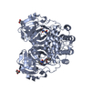
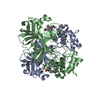
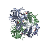
| Title | Crystal structure of MERS-CoV 3CL protease (C148A) in spacegroup P212121,pH 9.0 | ||||||
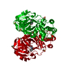
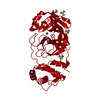
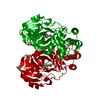
 Components Components | 3C-like proteinase | ||||||
 Keywords Keywords | VIRAL PROTEIN / MERS-CoV / 3CL protease | ||||||
| Function / homology |  Function and homology information Function and homology informationhost cell membrane / viral genome replication / methyltransferase activity / endonuclease activity / methylation / symbiont-mediated degradation of host mRNA / symbiont-mediated suppression of host ISG15-protein conjugation / G-quadruplex RNA binding / omega peptidase activity / symbiont-mediated perturbation of host ubiquitin-like protein modification ...host cell membrane / viral genome replication / methyltransferase activity / endonuclease activity / methylation / symbiont-mediated degradation of host mRNA / symbiont-mediated suppression of host ISG15-protein conjugation / G-quadruplex RNA binding / omega peptidase activity / symbiont-mediated perturbation of host ubiquitin-like protein modification / cysteine-type deubiquitinase activity / single-stranded RNA binding / viral protein processing / host cell perinuclear region of cytoplasm / symbiont-mediated suppression of host type I interferon-mediated signaling pathway / symbiont-mediated suppression of host gene expression / viral translational frameshifting / symbiont-mediated activation of host autophagy / cysteine-type endopeptidase activity / proteolysis / zinc ion binding Similarity search - Function | ||||||
| Biological species |  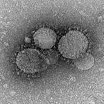 Middle East respiratory syndrome-related coronavirus Middle East respiratory syndrome-related coronavirus | ||||||
| Method |  X-RAY DIFFRACTION / SYNCHROTRON / MOLECULAR REPLACEMENT / Resolution: 2.78409685655 Å X-RAY DIFFRACTION / SYNCHROTRON / MOLECULAR REPLACEMENT / Resolution: 2.78409685655 Å | ||||||
 Authors Authors | Zhang, Y.T. / Zhou, X.L. / Li, J. / Zhang, J. | ||||||
 Citation Citation | Journal: To Be Published Title: Crystal structure of MERS-CoV 3CL protease (C148A) in spacegroup P212121,pH 9.0 Authors: Zhang, Y.T. / Zhou, X.L. / Li, J. / Zhang, J. | ||||||
| History |
|
- Structure visualization
| Structure viewer | Molecule: MolmilJmol/JSmol |
|---|
- Downloads & links
-Download
| PDBx/mmCIF format | 7dra.cif.gz | 139.3 KB | Display | PDBx/mmCIF format |
|---|---|---|---|---|
| PDB format | pdb7dra.ent.gz | 88.1 KB | Display | PDB format |
| PDBx/mmJSON format | 7dra.json.gz | Tree view | PDBx/mmJSON format | |
| Others |  Other downloads Other downloads |
-Validation report
| Arichive directory | https://data.pdbj.org/pub/pdb/validation_reports/dr/7draftp://data.pdbj.org/pub/pdb/validation_reports/dr/7dra | HTTPS FTP |
|---|
-Related structure data
| Related structure data |  7dr8S S: Starting model for refinement |
|---|---|
| Similar structure data |


-Links
 PDBj
PDBj
- Assembly
| Deposited unit | 
| ||||||||||||
|---|---|---|---|---|---|---|---|---|---|---|---|---|---|
| 1 |
| ||||||||||||
| Unit cell |
|
-Components
| #1: Protein | Mass: 33322.109 Da / Num. of mol.: 2 / Mutation: C148A Source method: isolated from a genetically manipulated source Source: (gene. exp.) Middle East respiratory syndrome-related coronavirusProduction host:  References: UniProt: T2B9I2, SARS coronavirus main proteinase |
|---|
-Experimental details
-Experiment
| Experiment | Method: X-RAY DIFFRACTION / Number of used crystals: 1 |
|---|
- Sample preparation
| Crystal | Density Matthews: 2.76 Å3/Da / Density % sol: 55.51 % |
|---|---|
| Crystal grow | Temperature: 298 K / Method: vapor diffusion, sitting drop Details: 4% 2-propanol, 0.1M BIS-TRIS Propane pH9.0 20% PEG5000 |
-Data collection
| Diffraction | Mean temperature: 100 K / Serial crystal experiment: N |
|---|---|
| Diffraction source | Source: SYNCHROTRON / Site: SSRF  / Beamline: BL17U1 / Wavelength: 0.97913 Å / Beamline: BL17U1 / Wavelength: 0.97913 Å |
| Detector | Type: ADSC QUANTUM 315r / Detector: CCD / Date: Nov 17, 2020 |
| Radiation | Protocol: SINGLE WAVELENGTH / Monochromatic (M) / Laue (L): M / Scattering type: x-ray |
| Radiation wavelength | Wavelength: 0.97913 Å / Relative weight: 1 |
| Reflection | Resolution: 2.78→51.76 Å / Num. obs: 18515 / % possible obs: 96.7 % / Redundancy: 13.2 % / Biso Wilson estimate: 58.3822186756 Å2 / Rpim(I) all: 0.048 / Net I/σ(I): 12.9 |
| Reflection shell | Resolution: 2.78→2.93 Å / Num. unique obs: 2739 / Rpim(I) all: 0.321 |
- Processing
| Software |
| ||||||||||||||||||||||||||||||||||||||||||||||||||||||||
|---|---|---|---|---|---|---|---|---|---|---|---|---|---|---|---|---|---|---|---|---|---|---|---|---|---|---|---|---|---|---|---|---|---|---|---|---|---|---|---|---|---|---|---|---|---|---|---|---|---|---|---|---|---|---|---|---|---|
| Refinement | Method to determine structure: MOLECULAR REPLACEMENT Starting model: 7DR8 Resolution: 2.78409685655→47.2425 Å / SU ML: 0.350511238448 / Cross valid method: NONE / σ(F): 1.34235233213 / Phase error: 27.0389175882 Stereochemistry target values: GeoStd + Monomer Library + CDL v1.2
| ||||||||||||||||||||||||||||||||||||||||||||||||||||||||
| Solvent computation | Shrinkage radii: 0.9 Å / VDW probe radii: 1.11 Å / Solvent model: FLAT BULK SOLVENT MODEL | ||||||||||||||||||||||||||||||||||||||||||||||||||||||||
| Displacement parameters | Biso mean: 50.9556542432 Å2 | ||||||||||||||||||||||||||||||||||||||||||||||||||||||||
| Refinement step | Cycle: LAST / Resolution: 2.78409685655→47.2425 Å
| ||||||||||||||||||||||||||||||||||||||||||||||||||||||||
| Refine LS restraints |
| ||||||||||||||||||||||||||||||||||||||||||||||||||||||||
| LS refinement shell |
|
