 Movie
Movie Controller


+ Open data

- Basic information
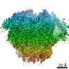
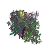
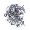
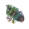
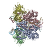
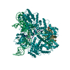
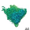
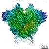
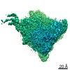
| Entry | Database: PDB / ID: 7dr1 | ||||||
|---|---|---|---|---|---|---|---|
| Title | Structure of Wild-type PSI monomer2 from Cyanophora paradoxa | ||||||
 Components Components |
| ||||||
 Keywords Keywords | PHOTOSYNTHESIS / Photosystem I / ELECTRON TRANSPORT | ||||||
| Function / homology |  Function and homology information Function and homology informationcyanelle thylakoid membrane / photosystem I reaction center / photosystem I / photosynthetic electron transport in photosystem I / photosystem I / chlorophyll binding / chloroplast thylakoid membrane / photosynthesis / 4 iron, 4 sulfur cluster binding / electron transfer activity ...cyanelle thylakoid membrane / photosystem I reaction center / photosystem I / photosynthetic electron transport in photosystem I / photosystem I / chlorophyll binding / chloroplast thylakoid membrane / photosynthesis / 4 iron, 4 sulfur cluster binding / electron transfer activity / oxidoreductase activity / magnesium ion binding / metal ion binding Similarity search - Function | ||||||
| Biological species |  Cyanophora paradoxa (eukaryote) Cyanophora paradoxa (eukaryote) | ||||||
| Method | ELECTRON MICROSCOPY / single particle reconstruction / cryo EM / Resolution: 3.2 Å | ||||||
 Authors Authors | Kato, K. / Nagao, R. / Akita, F. / Miyazaki, N. / Shen, J.R. | ||||||
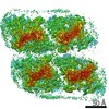
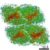
 Citation Citation | Journal: Nat Commun / Year: 2022 Title: Structure of a tetrameric photosystem I from a glaucophyte alga Cyanophora paradoxa. Authors: Koji Kato / Ryo Nagao / Yoshifumi Ueno / Makio Yokono / Takehiro Suzuki / Tian-Yi Jiang / Naoshi Dohmae / Fusamichi Akita / Seiji Akimoto / Naoyuki Miyazaki / Jian-Ren Shen /  Abstract: Photosystem I (PSI) is one of the two photosystems functioning in light-energy harvesting, transfer, and electron transfer in photosynthesis. However, the oligomerization state of PSI is variable ...Photosystem I (PSI) is one of the two photosystems functioning in light-energy harvesting, transfer, and electron transfer in photosynthesis. However, the oligomerization state of PSI is variable among photosynthetic organisms. We present a 3.8-Å resolution cryo-electron microscopic structure of tetrameric PSI isolated from the glaucophyte alga Cyanophora paradoxa, which reveals differences with PSI from other organisms in subunit composition and organization. The PSI tetramer is organized in a dimer of dimers with a C2 symmetry. Unlike cyanobacterial PSI tetramers, two of the four monomers are rotated around 90°, resulting in a completely different pattern of monomer-monomer interactions. Excitation-energy transfer among chlorophylls differs significantly between Cyanophora and cyanobacterial PSI tetramers. These structural and spectroscopic features reveal characteristic interactions and excitation-energy transfer in the Cyanophora PSI tetramer, suggesting that the Cyanophora PSI could represent a turning point in the evolution of PSI from prokaryotes to eukaryotes. #1: Journal: Biorxiv / Year: 2022Title: Structural insights into an evolutionary turning-point of photosystem I from prokaryotes to eukaryotes Authors: Kato, K. / Nagao, R. / Ueno, Y. / Yokono, M. / Suzuki, T. / Jiang, T.Y. / Dohmae, N. / Akita, F. / Akimoto, S. / Miyazaki, N. / Shen, J.R. | ||||||
| History |
|
- Structure visualization
| Movie |
Movie viewer |
|---|---|
| Structure viewer | Molecule: MolmilJmol/JSmol |
- Downloads & links
-Download
| PDBx/mmCIF format | 7dr1.cif.gz | 496.7 KB | Display | PDBx/mmCIF format |
|---|---|---|---|---|
| PDB format | pdb7dr1.ent.gz | 430.9 KB | Display | PDB format |
| PDBx/mmJSON format | 7dr1.json.gz | Tree view | PDBx/mmJSON format | |
| Others |  Other downloads Other downloads |
-Validation report
| Arichive directory | https://data.pdbj.org/pub/pdb/validation_reports/dr/7dr1ftp://data.pdbj.org/pub/pdb/validation_reports/dr/7dr1 | HTTPS FTP |
|---|
-Related structure data
| Related structure data | 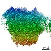 30821MC 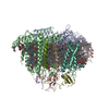 7dr0C  7dr2C M: map data used to model this data C: citing same article ( |
|---|---|
| Similar structure data |
-Links
 PDBj
PDBj


- Assembly
| Deposited unit | 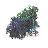
|
|---|---|
| 1 |
|
-Components
-Photosystem I P700 chlorophyll a apoprotein ... , 2 types, 2 molecules AB
| #1: Protein | Mass: 83397.305 Da / Num. of mol.: 1 / Source method: isolated from a natural source / Source: (natural) Cyanophora paradoxa (eukaryote) / References: UniProt: A0A097PBL3, photosystem I |
|---|---|
| #2: Protein | Mass: 82316.312 Da / Num. of mol.: 1 / Source method: isolated from a natural source / Source: (natural) Cyanophora paradoxa (eukaryote) / References: UniProt: P48113, photosystem I |
-Protein , 1 types, 1 molecules C
| #3: Protein | Mass: 8850.221 Da / Num. of mol.: 1 / Source method: isolated from a natural source / Source: (natural) Cyanophora paradoxa (eukaryote) / References: UniProt: P31173, photosystem I |
|---|
-Photosystem I reaction center subunit ... , 7 types, 7 molecules DEFIJLM
| #4: Protein | Mass: 23693.258 Da / Num. of mol.: 1 / Source method: isolated from a natural source / Source: (natural) Cyanophora paradoxa (eukaryote) / References: UniProt: Q9T4W8 |
|---|---|
| #5: Protein | Mass: 8058.164 Da / Num. of mol.: 1 / Source method: isolated from a natural source / Source: (natural) Cyanophora paradoxa (eukaryote) / References: UniProt: P48114 |
| #6: Protein | Mass: 20697.777 Da / Num. of mol.: 1 / Source method: isolated from a natural source / Source: (natural) Cyanophora paradoxa (eukaryote) / References: UniProt: P48115 |
| #7: Protein/peptide | Mass: 3772.620 Da / Num. of mol.: 1 / Source method: isolated from a natural source / Source: (natural) Cyanophora paradoxa (eukaryote) / References: UniProt: P48116 |
| #8: Protein/peptide | Mass: 4483.274 Da / Num. of mol.: 1 / Source method: isolated from a natural source / Source: (natural) Cyanophora paradoxa (eukaryote) / References: UniProt: P48117 |
| #9: Protein | Mass: 15376.591 Da / Num. of mol.: 1 / Source method: isolated from a natural source / Source: (natural) Cyanophora paradoxa (eukaryote) |
| #10: Protein/peptide | Mass: 3334.065 Da / Num. of mol.: 1 / Source method: isolated from a natural source / Source: (natural) Cyanophora paradoxa (eukaryote) / References: UniProt: P48185 |
-Non-polymers , 7 types, 108 molecules 


| #11: Chemical | ChemComp-CL0 /  Mass: 893.489 Da / Num. of mol.: 1 / Source method: obtained synthetically / Formula: C55H72MgN4O5 Mass: 893.489 Da / Num. of mol.: 1 / Source method: obtained synthetically / Formula: C55H72MgN4O5 | ||||||||||
|---|---|---|---|---|---|---|---|---|---|---|---|
| #12: Chemical | ChemComp-CLA /  Mass: 893.489 Da / Num. of mol.: 80 / Source method: obtained synthetically / Formula: C55H72MgN4O5 Mass: 893.489 Da / Num. of mol.: 80 / Source method: obtained synthetically / Formula: C55H72MgN4O5#13: Chemical |  Mass: 450.696 Da / Num. of mol.: 2 / Source method: obtained synthetically / Formula: C31H46O2 Mass: 450.696 Da / Num. of mol.: 2 / Source method: obtained synthetically / Formula: C31H46O2#14: Chemical |  Mass: 351.640 Da / Num. of mol.: 3 / Source method: obtained synthetically / Formula: Fe4S4 Mass: 351.640 Da / Num. of mol.: 3 / Source method: obtained synthetically / Formula: Fe4S4#15: Chemical | ChemComp-BCR /  Mass: 536.873 Da / Num. of mol.: 19 / Source method: obtained synthetically / Formula: C40H56 Mass: 536.873 Da / Num. of mol.: 19 / Source method: obtained synthetically / Formula: C40H56#16: Chemical |  Mass: 722.970 Da / Num. of mol.: 2 / Source method: obtained synthetically / Formula: C38H75O10P / Comment: phospholipid*YM Mass: 722.970 Da / Num. of mol.: 2 / Source method: obtained synthetically / Formula: C38H75O10P / Comment: phospholipid*YM#17: Chemical | ChemComp-LMG / |  Mass: 787.158 Da / Num. of mol.: 1 / Source method: obtained synthetically / Formula: C45H86O10 Mass: 787.158 Da / Num. of mol.: 1 / Source method: obtained synthetically / Formula: C45H86O10 |
-Details
| Has ligand of interest | N |
|---|---|
| Has protein modification | Y |
-Experimental details
-Experiment
| Experiment | Method: ELECTRON MICROSCOPY |
|---|---|
| EM experiment | Aggregation state: PARTICLE / 3D reconstruction method: single particle reconstruction |
- Sample preparation
| Component | Name: PSI monomer2 / Type: COMPLEX / Entity ID: #1-#10 / Source: NATURAL | ||||||||||||
|---|---|---|---|---|---|---|---|---|---|---|---|---|---|
| Molecular weight | Value: 0.35 MDa / Experimental value: NO | ||||||||||||
| Source (natural) | Organism: Cyanophora paradoxa (eukaryote) | ||||||||||||
| Buffer solution | pH: 6.5 | ||||||||||||
| Buffer component |
| ||||||||||||
| Specimen | Conc.: 0.007 mg/ml / Embedding applied: NO / Shadowing applied: NO / Staining applied: NO / Vitrification applied: YES | ||||||||||||
| Specimen support | Grid material: COPPER / Grid mesh size: 300 divisions/in. / Grid type: Quantifoil R2/1 | ||||||||||||
| Vitrification | Instrument: FEI VITROBOT MARK IV / Cryogen name: ETHANE / Humidity: 100 % / Chamber temperature: 277 K |
- Electron microscopy imaging
| Experimental equipment |  Model: Talos Arctica / Image courtesy: FEI Company |
|---|---|
| Microscopy | Model: FEI TALOS ARCTICA |
| Electron gun | Electron source:  FIELD EMISSION GUN / Accelerating voltage: 200 kV / Illumination mode: FLOOD BEAM FIELD EMISSION GUN / Accelerating voltage: 200 kV / Illumination mode: FLOOD BEAM |
| Electron lens | Mode: BRIGHT FIELD |
| Image recording | Electron dose: 50 e/Å2 / Film or detector model: FEI FALCON III (4k x 4k) |
- Processing
| EM software |
| ||||||||||||||||||||||||||||||||||||
|---|---|---|---|---|---|---|---|---|---|---|---|---|---|---|---|---|---|---|---|---|---|---|---|---|---|---|---|---|---|---|---|---|---|---|---|---|---|
| CTF correction | Type: PHASE FLIPPING AND AMPLITUDE CORRECTION | ||||||||||||||||||||||||||||||||||||
| Particle selection | Num. of particles selected: 1603082 | ||||||||||||||||||||||||||||||||||||
| Symmetry | Point symmetry: C1 (asymmetric) | ||||||||||||||||||||||||||||||||||||
| 3D reconstruction | Resolution: 3.2 Å / Resolution method: FSC 0.143 CUT-OFF / Num. of particles: 110380 / Algorithm: FOURIER SPACE / Symmetry type: POINT | ||||||||||||||||||||||||||||||||||||
| Atomic model building | Protocol: FLEXIBLE FIT / Space: REAL / Target criteria: Correlation coefficient |
