 Movie
Movie Controller


+ Open data

- Basic information
| Entry | Database: PDB / ID: 7dov | ||||||
|---|---|---|---|---|---|---|---|
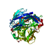
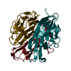
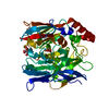
| Title | The crystal structure of zebrafish tumor necrosis factor alpha | ||||||
 Components Components | Lymphotoxin-alpha | ||||||
 Keywords Keywords | CYTOKINE / TNF alpha / Danio rerio / Zebrafish | ||||||
| Function / homology |  Function and homology information Function and homology informationfin regeneration / regeneration / intestinal epithelial structure maintenance / response to arsenic-containing substance / tumor necrosis factor receptor binding / response to exogenous dsRNA / membrane => GO:0016020 / response to bacterium / liver development / response to molecule of bacterial origin ...fin regeneration / regeneration / intestinal epithelial structure maintenance / response to arsenic-containing substance / tumor necrosis factor receptor binding / response to exogenous dsRNA / membrane => GO:0016020 / response to bacterium / liver development / response to molecule of bacterial origin / autophagy / response to virus / response to lipopolysaccharide / defense response to bacterium / immune response / inflammatory response Similarity search - Function | ||||||
| Biological species |  | ||||||
| Method |  X-RAY DIFFRACTION / SYNCHROTRON / MOLECULAR REPLACEMENT / Resolution: 2.59 Å X-RAY DIFFRACTION / SYNCHROTRON / MOLECULAR REPLACEMENT / Resolution: 2.59 Å | ||||||
 Authors Authors | Duan, Y. / Xia, C. | ||||||
| Funding support |  China, 1items China, 1items
| ||||||
 Citation Citation | Journal: To Be Published Title: The structure of zebrafish tumour necrosis factor alpha at 2.6 angstrom resolution Authors: Duan, Y. / Xia, C. | ||||||
| History |
|
- Structure visualization
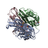
| Structure viewer | Molecule: MolmilJmol/JSmol |
|---|
- Downloads & links
-Download
| PDBx/mmCIF format | 7dov.cif.gz | 102.6 KB | Display | PDBx/mmCIF format |
|---|---|---|---|---|
| PDB format | pdb7dov.ent.gz | 77.4 KB | Display | PDB format |
| PDBx/mmJSON format | 7dov.json.gz | Tree view | PDBx/mmJSON format | |
| Others |  Other downloads Other downloads |
-Validation report
| Arichive directory | https://data.pdbj.org/pub/pdb/validation_reports/do/7dovftp://data.pdbj.org/pub/pdb/validation_reports/do/7dov | HTTPS FTP |
|---|
-Related structure data
| Related structure data |  4tsvS S: Starting model for refinement |
|---|---|
| Similar structure data |


-Links
 PDBj
PDBj- Assembly
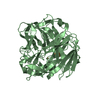
| Deposited unit | 
| ||||||||||||
|---|---|---|---|---|---|---|---|---|---|---|---|---|---|
| 1 |
| ||||||||||||
| Unit cell |
|
-Components
| #1: Protein | Mass: 17273.205 Da / Num. of mol.: 3 Source method: isolated from a genetically manipulated source Source: (gene. exp.)  #2: Water | ChemComp-HOH / |  Mass: 18.015 Da / Num. of mol.: 69 / Source method: isolated from a natural source / Formula: H2O Mass: 18.015 Da / Num. of mol.: 69 / Source method: isolated from a natural source / Formula: H2OHas protein modification | Y | |
|---|
-Experimental details
-Experiment
| Experiment | Method: X-RAY DIFFRACTION / Number of used crystals: 1 |
|---|
- Sample preparation
| Crystal | Density Matthews: 2.35 Å3/Da / Density % sol: 47.61 % |
|---|---|
| Crystal grow | Temperature: 277.15 K / Method: vapor diffusion, sitting drop / pH: 7 Details: 0.1 M DL-Malic acid pH 7.0, 12% w/v Polyethylene glycol 3350 |
-Data collection
| Diffraction | Mean temperature: 100 K / Serial crystal experiment: N |
|---|---|
| Diffraction source | Source: SYNCHROTRON / Site: SSRF / Beamline: BL19U1 / Wavelength: 0.97916 Å |
| Detector | Type: ADSC QUANTUM 315r / Detector: CCD / Date: Oct 26, 2019 |
| Radiation | Protocol: SINGLE WAVELENGTH / Monochromatic (M) / Laue (L): M / Scattering type: x-ray |
| Radiation wavelength | Wavelength: 0.97916 Å / Relative weight: 1 |
| Reflection | Resolution: 2.59→51.31 Å / Num. obs: 15728 / % possible obs: 100 % / Redundancy: 25.1 % / Biso Wilson estimate: 36.59 Å2 / Rmerge(I) obs: 0.096 / Net I/σ(I): 32.3 |
| Reflection shell | Resolution: 2.6→2.64 Å / Rmerge(I) obs: 0.852 / Num. unique obs: 1428 |
- Processing
| Software |
| ||||||||||||||||||||||||||||||||||||||||||
|---|---|---|---|---|---|---|---|---|---|---|---|---|---|---|---|---|---|---|---|---|---|---|---|---|---|---|---|---|---|---|---|---|---|---|---|---|---|---|---|---|---|---|---|
| Refinement | Method to determine structure: MOLECULAR REPLACEMENT Starting model: 4TSV Resolution: 2.59→44.27 Å / SU ML: 0.3566 / Cross valid method: FREE R-VALUE / σ(F): 1.37 / Phase error: 29.7533 Stereochemistry target values: GeoStd + Monomer Library + CDL v1.2
| ||||||||||||||||||||||||||||||||||||||||||
| Solvent computation | Shrinkage radii: 0.9 Å / VDW probe radii: 1.11 Å / Solvent model: FLAT BULK SOLVENT MODEL | ||||||||||||||||||||||||||||||||||||||||||
| Displacement parameters | Biso mean: 50.13 Å2 | ||||||||||||||||||||||||||||||||||||||||||
| Refinement step | Cycle: LAST / Resolution: 2.59→44.27 Å
| ||||||||||||||||||||||||||||||||||||||||||
| Refine LS restraints |
| ||||||||||||||||||||||||||||||||||||||||||
| LS refinement shell |
|
