 Movie
Movie Controller


+ Open data

- Basic information
| Entry | Database: PDB / ID: 7doq | ||||||
|---|---|---|---|---|---|---|---|
| Title | Lp major histidine acid phosphatase mutant D281A/5'-AMP | ||||||
 Components Components | Acid phosphatase | ||||||
 Keywords Keywords | HYDROLASE / Complex / 5'-AMP | ||||||
| Function / homology | Histidine acid phosphatases phosphohistidine signature. / Histidine acid phosphatase active site / Histidine phosphatase superfamily, clade-2 / Histidine phosphatase superfamily (branch 2) / Histidine phosphatase superfamily / PHOSPHATE ION / Acid phosphatase Function and homology information Function and homology information | ||||||
| Biological species |   Legionella pneumophila (bacteria) Legionella pneumophila (bacteria) | ||||||
| Method |  X-RAY DIFFRACTION / SYNCHROTRON / MOLECULAR REPLACEMENT / Resolution: 2.2 Å X-RAY DIFFRACTION / SYNCHROTRON / MOLECULAR REPLACEMENT / Resolution: 2.2 Å | ||||||
 Authors Authors | Guo, Y. / Teng, Y. | ||||||
| Funding support |  China, 1items China, 1items
| ||||||
 Citation Citation | Journal: Biochem.Biophys.Res.Commun. / Year: 2021 Title: Structural insights into a new substrate binding mode of a histidine acid phosphatase from Legionella pneumophila. Authors: Guo, Y. / Zhou, D. / Zhang, H. / Zhang, N.N. / Qi, X. / Chen, X. / Chen, Q. / Li, J. / Ge, H. / Teng, Y.B. | ||||||
| History |
|
- Structure visualization
| Structure viewer | Molecule: MolmilJmol/JSmol |
|---|
- Downloads & links
-Download
| PDBx/mmCIF format | 7doq.cif.gz | 271.3 KB | Display | PDBx/mmCIF format |
|---|---|---|---|---|
| PDB format | pdb7doq.ent.gz | 220.3 KB | Display | PDB format |
| PDBx/mmJSON format | 7doq.json.gz | Tree view | PDBx/mmJSON format | |
| Others |  Other downloads Other downloads |
-Validation report
| Summary document | 7doq_validation.pdf.gz | 470.8 KB | Display | wwPDB validaton report |
|---|---|---|---|---|
| Full document | 7doq_full_validation.pdf.gz | 487.4 KB | Display | |
| Data in XML | 7doq_validation.xml.gz | 49 KB | Display | |
| Data in CIF | 7doq_validation.cif.gz | 67.6 KB | Display | |
| Arichive directory | https://data.pdbj.org/pub/pdb/validation_reports/do/7doqftp://data.pdbj.org/pub/pdb/validation_reports/do/7doq | HTTPS FTP |
-Related structure data
| Related structure data |  7d2fC  3it0S S: Starting model for refinement C: citing same article ( |
|---|---|
| Similar structure data |


-Links
 PDBj
PDBj


- Assembly
| Deposited unit | 
| ||||||||
|---|---|---|---|---|---|---|---|---|---|
| 1 | 
| ||||||||
| 2 | 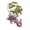
| ||||||||
| Unit cell |
|
-Components
| #1: Protein | Mass: 37705.602 Da / Num. of mol.: 4 Source method: isolated from a genetically manipulated source Source: (gene. exp.) Legionella pneumophila (bacteria) / Gene: C3927_05010, DI026_02700 / Production host: #2: Chemical | ChemComp-PO4 /   Mass: 94.971 Da / Num. of mol.: 7 / Source method: obtained synthetically / Formula: PO4 Mass: 94.971 Da / Num. of mol.: 7 / Source method: obtained synthetically / Formula: PO4#3: Water | ChemComp-HOH / |  Mass: 18.015 Da / Num. of mol.: 259 / Source method: isolated from a natural source / Formula: H2O Mass: 18.015 Da / Num. of mol.: 259 / Source method: isolated from a natural source / Formula: H2OHas ligand of interest | N | Has protein modification | Y | |
|---|
-Experimental details
-Experiment
| Experiment | Method: X-RAY DIFFRACTION / Number of used crystals: 1 |
|---|
- Sample preparation
| Crystal | Density Matthews: 2.38 Å3/Da / Density % sol: 48.35 % |
|---|---|
| Crystal grow | Temperature: 289.15 K / Method: vapor diffusion, sitting drop Details: 20% w/v Polyethylene glycol 3350, 0.2M Sodium formate, 0.025% (w/v) low gelling temperature agarose |
-Data collection
| Diffraction | Mean temperature: 298.15 K / Serial crystal experiment: N |
|---|---|
| Diffraction source | Source: SYNCHROTRON / Site: SSRF / Beamline: BL17U / Wavelength: 0.978 Å |
| Detector | Type: MARRESEARCH / Detector: CCD / Date: Dec 17, 2016 |
| Radiation | Monochromator: 0.978 / Protocol: SINGLE WAVELENGTH / Monochromatic (M) / Laue (L): M / Scattering type: x-ray |
| Radiation wavelength | Wavelength: 0.978 Å / Relative weight: 1 |
| Reflection | Resolution: 2.2→35.62 Å / Num. obs: 66012 / % possible obs: 96.37 % / Redundancy: 2.2 % / Rmerge(I) obs: 0.082 / Net I/σ(I): 11.6 |
| Reflection shell | Resolution: 2.2→2.255 Å / Rmerge(I) obs: 0.479 / Num. unique obs: 4815 |
- Processing
| Software |
| ||||||||||||||||||||||||||||||||||||||||||||||||||||||||||||
|---|---|---|---|---|---|---|---|---|---|---|---|---|---|---|---|---|---|---|---|---|---|---|---|---|---|---|---|---|---|---|---|---|---|---|---|---|---|---|---|---|---|---|---|---|---|---|---|---|---|---|---|---|---|---|---|---|---|---|---|---|---|
| Refinement | Method to determine structure: MOLECULAR REPLACEMENT Starting model: 3IT0 Resolution: 2.2→35.62 Å / Cor.coef. Fo:Fc: 0.955 / Cor.coef. Fo:Fc free: 0.931 / Cross valid method: THROUGHOUT / σ(F): 0 / ESU R: 0.3 / ESU R Free: 0.21 / Stereochemistry target values: MAXIMUM LIKELIHOOD Details: HYDROGENS HAVE BEEN ADDED IN THE RIDING POSITIONS U VALUES : REFINED INDIVIDUALLY
| ||||||||||||||||||||||||||||||||||||||||||||||||||||||||||||
| Solvent computation | Ion probe radii: 0.8 Å / Shrinkage radii: 0.8 Å / VDW probe radii: 1.2 Å / Solvent model: MASK | ||||||||||||||||||||||||||||||||||||||||||||||||||||||||||||
| Displacement parameters | Biso max: 258.21 Å2 / Biso mean: 40.252 Å2 / Biso min: 18.51 Å2
| ||||||||||||||||||||||||||||||||||||||||||||||||||||||||||||
| Refinement step | Cycle: final / Resolution: 2.2→35.62 Å
| ||||||||||||||||||||||||||||||||||||||||||||||||||||||||||||
| Refine LS restraints |
| ||||||||||||||||||||||||||||||||||||||||||||||||||||||||||||
| LS refinement shell | Resolution: 2.2→2.255 Å / Rfactor Rfree error: 0
|
