 Movie
Movie Controller

[English] 日本語
 Yorodumi
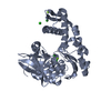
- PDB-7dmw: Crystal structure of CcpC regulatory domain in complex with citra... -
+ Open data

- Basic information
| Entry | Database: PDB / ID: 7dmw | |||||||||
|---|---|---|---|---|---|---|---|---|---|---|
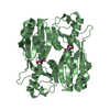
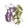
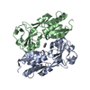
| Title | Crystal structure of CcpC regulatory domain in complex with citrate from Bacillus amyloliquefaciens | |||||||||
 Components Components | CcpC | |||||||||
 Keywords Keywords | TRANSCRIPTION / CcpC / Complex / Bacillus amyloliquefaciens / Transcriptional regulator / citrate | |||||||||
| Function / homology |  Function and homology information Function and homology informationtranscription cis-regulatory region binding / DNA-binding transcription factor activity / DNA-templated transcription Similarity search - Function | |||||||||
| Biological species |  | |||||||||
| Method |  X-RAY DIFFRACTION / SYNCHROTRON / SAD / Resolution: 2.29 Å X-RAY DIFFRACTION / SYNCHROTRON / SAD / Resolution: 2.29 Å | |||||||||
 Authors Authors | Chen, J. / Wang, L. / Shang, F. / Liu, W. / Chen, Y. / Lan, J. / Bu, T. / Bai, X. / Xu, Y. | |||||||||
| Funding support |  China, 2items China, 2items
| |||||||||
 Citation Citation | Journal: Sci Rep / Year: 2021 Title: Functional and structural analysis of catabolite control protein C that responds to citrate. Authors: Liu, W. / Chen, J. / Jin, L. / Liu, Z.Y. / Lu, M. / Jiang, G. / Yang, Q. / Quan, C. / Nam, K.H. / Xu, Y. | |||||||||
| History |
|
- Structure visualization
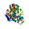
| Structure viewer | Molecule: MolmilJmol/JSmol |
|---|
- Downloads & links
-Download
| PDBx/mmCIF format | 7dmw.cif.gz | 263.5 KB | Display | PDBx/mmCIF format |
|---|---|---|---|---|
| PDB format | pdb7dmw.ent.gz | 171.4 KB | Display | PDB format |
| PDBx/mmJSON format | 7dmw.json.gz | Tree view | PDBx/mmJSON format | |
| Others |  Other downloads Other downloads |
-Validation report
| Arichive directory | https://data.pdbj.org/pub/pdb/validation_reports/dm/7dmwftp://data.pdbj.org/pub/pdb/validation_reports/dm/7dmw | HTTPS FTP |
|---|
-Related structure data
| Similar structure data |
|---|


-Links
 PDBj
PDBj


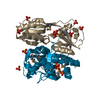
- Assembly
| Deposited unit | 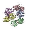
| ||||||||||||
|---|---|---|---|---|---|---|---|---|---|---|---|---|---|
| 1 | 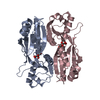
| ||||||||||||
| 2 | 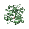
| ||||||||||||
| 3 | 
| ||||||||||||
| Unit cell |
|
-Components
| #1: Protein | Mass: 34031.148 Da / Num. of mol.: 5 Source method: isolated from a genetically manipulated source Source: (gene. exp.) #2: Chemical | ChemComp-FLC /   Mass: 189.100 Da / Num. of mol.: 5 / Source method: isolated from a natural source / Formula: C6H5O7 / Feature type: SUBJECT OF INVESTIGATION Mass: 189.100 Da / Num. of mol.: 5 / Source method: isolated from a natural source / Formula: C6H5O7 / Feature type: SUBJECT OF INVESTIGATION#3: Water | ChemComp-HOH / |  Mass: 18.015 Da / Num. of mol.: 153 / Source method: isolated from a natural source / Formula: H2O Mass: 18.015 Da / Num. of mol.: 153 / Source method: isolated from a natural source / Formula: H2OHas ligand of interest | Y | |
|---|
-Experimental details
-Experiment
| Experiment | Method: X-RAY DIFFRACTION / Number of used crystals: 1 |
|---|
- Sample preparation
| Crystal | Density Matthews: 1.91 Å3/Da / Density % sol: 35.53 % |
|---|---|
| Crystal grow | Temperature: 287 K / Method: vapor diffusion, sitting drop / pH: 7.5 / Details: 10% PEG 6000 5% MPD 0.1 M HEPES (pH7.5) / PH range: 6.5-7.5 |
-Data collection
| Diffraction | Mean temperature: 100 K / Ambient temp details: Liquid nitrogen / Serial crystal experiment: N |
|---|---|
| Diffraction source | Source: SYNCHROTRON / Site: PAL/PLS  / Beamline: 7A (6B, 6C1) / Wavelength: 0.9826 Å / Beamline: 7A (6B, 6C1) / Wavelength: 0.9826 Å |
| Detector | Type: ADSC QUANTUM 315r / Detector: CCD / Date: Nov 15, 2017 |
| Radiation | Protocol: SINGLE WAVELENGTH / Monochromatic (M) / Laue (L): M / Scattering type: x-ray |
| Radiation wavelength | Wavelength: 0.9826 Å / Relative weight: 1 |
| Reflection | Resolution: 2.29→50 Å / Num. obs: 55746 / % possible obs: 96.53 % / Redundancy: 5.3 % / Biso Wilson estimate: 48.74 Å2 / Rsym value: 0.08 / Net I/σ(I): 36.2 |
| Reflection shell | Resolution: 2.3→2.34 Å / Redundancy: 2.6 % / Mean I/σ(I) obs: 2.64 / Num. unique obs: 2427 / Rsym value: 0.31 / % possible all: 84.1 |
- Processing
| Software |
| |||||||||||||||||||||||||||||||||||||||||||||||||||||||||||||||||||||||||||||||||||||||||||||||||||||||||
|---|---|---|---|---|---|---|---|---|---|---|---|---|---|---|---|---|---|---|---|---|---|---|---|---|---|---|---|---|---|---|---|---|---|---|---|---|---|---|---|---|---|---|---|---|---|---|---|---|---|---|---|---|---|---|---|---|---|---|---|---|---|---|---|---|---|---|---|---|---|---|---|---|---|---|---|---|---|---|---|---|---|---|---|---|---|---|---|---|---|---|---|---|---|---|---|---|---|---|---|---|---|---|---|---|---|---|
| Refinement | Method to determine structure: SAD / Resolution: 2.29→29.71 Å / SU ML: 0.3891 / Cross valid method: FREE R-VALUE / σ(F): 1.18 / Phase error: 32.379 Stereochemistry target values: GeoStd + Monomer Library + CDL v1.2
| |||||||||||||||||||||||||||||||||||||||||||||||||||||||||||||||||||||||||||||||||||||||||||||||||||||||||
| Solvent computation | Shrinkage radii: 0.9 Å / VDW probe radii: 1.11 Å / Solvent model: FLAT BULK SOLVENT MODEL | |||||||||||||||||||||||||||||||||||||||||||||||||||||||||||||||||||||||||||||||||||||||||||||||||||||||||
| Displacement parameters | Biso mean: 62 Å2 | |||||||||||||||||||||||||||||||||||||||||||||||||||||||||||||||||||||||||||||||||||||||||||||||||||||||||
| Refinement step | Cycle: LAST / Resolution: 2.29→29.71 Å
| |||||||||||||||||||||||||||||||||||||||||||||||||||||||||||||||||||||||||||||||||||||||||||||||||||||||||
| Refine LS restraints |
| |||||||||||||||||||||||||||||||||||||||||||||||||||||||||||||||||||||||||||||||||||||||||||||||||||||||||
| LS refinement shell |
|
