 Movie
Movie Controller


+ Open data

- Basic information
| Entry | Database: PDB / ID: 7dkr | |||||||||
|---|---|---|---|---|---|---|---|---|---|---|
| Title | Crystal structure of native E. coli Grx2 at 2.38 A | |||||||||
 Components Components | Glutaredoxin | |||||||||
 Keywords Keywords | OXIDOREDUCTASE / E. coli Grx2 / Native | |||||||||
| Function / homology |  Function and homology information Function and homology information | |||||||||
| Biological species |  | |||||||||
| Method |  X-RAY DIFFRACTION / SYNCHROTRON / MOLECULAR REPLACEMENT / Resolution: 2.378 Å X-RAY DIFFRACTION / SYNCHROTRON / MOLECULAR REPLACEMENT / Resolution: 2.378 Å | |||||||||
 Authors Authors | Sreekumar, S.N. / Arockiasamy, A. | |||||||||
| Funding support |  India, 2items India, 2items
| |||||||||
 Citation Citation | Journal: To Be Published Title: Crystal structure of native E. coli Grx2 at 2.38 A Authors: Sreekumar, S.N. / Arockiasamy, A. | |||||||||
| History |
|
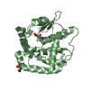
- Structure visualization
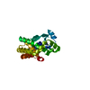
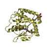
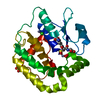
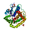
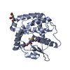
| Structure viewer | Molecule: MolmilJmol/JSmol |
|---|
- Downloads & links
-Download
| PDBx/mmCIF format | 7dkr.cif.gz | 54.7 KB | Display | PDBx/mmCIF format |
|---|---|---|---|---|
| PDB format | pdb7dkr.ent.gz | 37.8 KB | Display | PDB format |
| PDBx/mmJSON format | 7dkr.json.gz | Tree view | PDBx/mmJSON format | |
| Others |  Other downloads Other downloads |
-Validation report
| Summary document | 7dkr_validation.pdf.gz | 422.5 KB | Display | wwPDB validaton report |
|---|---|---|---|---|
| Full document | 7dkr_full_validation.pdf.gz | 423.6 KB | Display | |
| Data in XML | 7dkr_validation.xml.gz | 9.1 KB | Display | |
| Data in CIF | 7dkr_validation.cif.gz | 11.2 KB | Display | |
| Arichive directory | https://data.pdbj.org/pub/pdb/validation_reports/dk/7dkrftp://data.pdbj.org/pub/pdb/validation_reports/dk/7dkr | HTTPS FTP |
-Related structure data
| Related structure data | 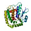 4kx4S S: Starting model for refinement |
|---|---|
| Similar structure data |

-Links
 PDBj
PDBj

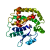
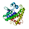
- Assembly
| Deposited unit | 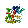
| ||||||||
|---|---|---|---|---|---|---|---|---|---|
| 1 |
| ||||||||
| Unit cell |
|
-Components
| #1: Protein | Mass: 24383.229 Da / Num. of mol.: 1 Source method: isolated from a genetically manipulated source Source: (gene. exp.) |
|---|
-Experimental details
-Experiment
| Experiment | Method: X-RAY DIFFRACTION / Number of used crystals: 1 |
|---|
- Sample preparation
| Crystal | Density Matthews: 2.06 Å3/Da / Density % sol: 39.9 % / Description: 2D Crystals |
|---|---|
| Crystal grow | Temperature: 293.15 K / Method: vapor diffusion, sitting drop / pH: 7.5 Details: Protein solution: 25mg/ml in 25mM Tris pH 8.0, 150 mM NaCl, Reservoir condition:0.1M HEPES pH:7.5, 20% w/v PEG 10000, Protein/reservoir mixed in 1:1, 1:2 and 2:1 ratio, set up using MRC ...Details: Protein solution: 25mg/ml in 25mM Tris pH 8.0, 150 mM NaCl, Reservoir condition:0.1M HEPES pH:7.5, 20% w/v PEG 10000, Protein/reservoir mixed in 1:1, 1:2 and 2:1 ratio, set up using MRC Swissci 3 well plates with the drop size of 150 nl |
-Data collection
| Diffraction | Mean temperature: 100 K / Serial crystal experiment: N |
|---|---|
| Diffraction source | Source: SYNCHROTRON / Site: ELETTRA  / Beamline: 11.2C / Wavelength: 1 Å / Beamline: 11.2C / Wavelength: 1 Å |
| Detector | Type: DECTRIS PILATUS 6M / Detector: PIXEL / Date: Feb 12, 2020 |
| Radiation | Protocol: SINGLE WAVELENGTH / Monochromatic (M) / Laue (L): M / Scattering type: x-ray |
| Radiation wavelength | Wavelength: 1 Å / Relative weight: 1 |
| Reflection | Resolution: 2.38→89.28 Å / Num. obs: 8647 / % possible obs: 100 % / Redundancy: 11 % / Biso Wilson estimate: 19 Å2 / CC1/2: 0.997 / Rmerge(I) obs: 0.115 / Rpim(I) all: 0.036 / Rrim(I) all: 0.12 / Χ2: 0.92 / Net I/σ(I): 15.2 |
| Reflection shell | Resolution: 2.38→2.47 Å / Redundancy: 9.5 % / Rmerge(I) obs: 0.314 / Mean I/σ(I) obs: 6.5 / Num. unique obs: 877 / CC1/2: 0.973 / Rpim(I) all: 0.106 / Rrim(I) all: 0.332 / Χ2: 0.89 / % possible all: 100 |
- Processing
| Software |
| ||||||||||||||||||
|---|---|---|---|---|---|---|---|---|---|---|---|---|---|---|---|---|---|---|---|
| Refinement | Method to determine structure: MOLECULAR REPLACEMENT Starting model: 4KX4 Resolution: 2.378→39.475 Å / SU ML: 0.27 / Cross valid method: THROUGHOUT / σ(F): 1.37 / Phase error: 23.51 / Stereochemistry target values: ML
| ||||||||||||||||||
| Solvent computation | Shrinkage radii: 0.9 Å / VDW probe radii: 1.11 Å / Solvent model: FLAT BULK SOLVENT MODEL | ||||||||||||||||||
| Displacement parameters | Biso max: 73.34 Å2 / Biso mean: 18.5777 Å2 / Biso min: 5.96 Å2 | ||||||||||||||||||
| Refinement step | Cycle: final / Resolution: 2.378→39.475 Å
| ||||||||||||||||||
| LS refinement shell | Refine-ID: X-RAY DIFFRACTION / Rfactor Rfree error: 0 / % reflection obs: 100 %
|
