 Movie
Movie Controller

[English] 日本語
 Yorodumi
- PDB-7d9l: Crystal structure of E. coli Grx2: Enzyme inhibited state in comp... -
+ Open data

- Basic information
| Entry | Database: PDB / ID: 7d9l | |||||||||
|---|---|---|---|---|---|---|---|---|---|---|
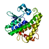
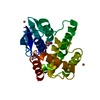
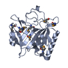
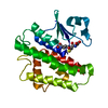
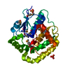
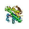
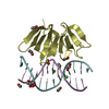
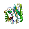
| Title | Crystal structure of E. coli Grx2: Enzyme inhibited state in complex with Zinc and glutathione sulfinic acid | |||||||||
 Components Components | Glutaredoxin | |||||||||
 Keywords Keywords | OXIDOREDUCTASE / E. coli / GRX2 / Zinc / Glutathione sulfinic acid / Enzyme inhibition | |||||||||
| Function / homology |  Function and homology information Function and homology information | |||||||||
| Biological species |  | |||||||||
| Method |  X-RAY DIFFRACTION / SYNCHROTRON / MOLECULAR REPLACEMENT / Resolution: 1.61 Å X-RAY DIFFRACTION / SYNCHROTRON / MOLECULAR REPLACEMENT / Resolution: 1.61 Å | |||||||||
 Authors Authors | Sreekumar, S.N. / Arockiasamy, A. | |||||||||
| Funding support |  India, 2items India, 2items
| |||||||||
 Citation Citation | Journal: To Be Published Title: Crystal structure of E. coli Grx2: Enzyme inhibited state in complex with Zinc and glutathione sulfinic acid Authors: Sreekumar, S.N. / Arockiasamy, A. #1: Journal: J Mol Biol / Year: 2001Title: Solution structure of Escherichia coli glutaredoxin-2 shows similarity to mammalian glutathione-S-transferases. Authors: Xia, B. / Vlamis-Gardikas, A. / Holmgren, A. / Wright, P.E. / Dyson, H.J. | |||||||||
| History |
|
- Structure visualization
| Structure viewer | Molecule: MolmilJmol/JSmol |
|---|
- Downloads & links
-Download
| PDBx/mmCIF format | 7d9l.cif.gz | 108.6 KB | Display | PDBx/mmCIF format |
|---|---|---|---|---|
| PDB format | pdb7d9l.ent.gz | 80.8 KB | Display | PDB format |
| PDBx/mmJSON format | 7d9l.json.gz | Tree view | PDBx/mmJSON format | |
| Others |  Other downloads Other downloads |
-Validation report
| Arichive directory | https://data.pdbj.org/pub/pdb/validation_reports/d9/7d9lftp://data.pdbj.org/pub/pdb/validation_reports/d9/7d9l | HTTPS FTP |
|---|
-Related structure data
| Related structure data | 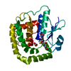 4kx4S S: Starting model for refinement |
|---|---|
| Similar structure data |


-Links
 PDBj
PDBj


- Assembly
| Deposited unit | 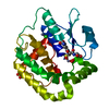
| ||||||||
|---|---|---|---|---|---|---|---|---|---|
| 1 |
| ||||||||
| Unit cell |
|
-Components
-Protein , 1 types, 1 molecules A
| #1: Protein | Mass: 24383.229 Da / Num. of mol.: 1 Source method: isolated from a genetically manipulated source Source: (gene. exp.) |
|---|
-Non-polymers , 6 types, 162 molecules 


| #2: Chemical |  Mass: 65.409 Da / Num. of mol.: 2 / Source method: obtained synthetically / Formula: Zn / Feature type: SUBJECT OF INVESTIGATION Mass: 65.409 Da / Num. of mol.: 2 / Source method: obtained synthetically / Formula: Zn / Feature type: SUBJECT OF INVESTIGATION#3: Chemical | ChemComp-CL / |  Mass: 35.453 Da / Num. of mol.: 1 / Source method: obtained synthetically / Formula: Cl / Feature type: SUBJECT OF INVESTIGATION Mass: 35.453 Da / Num. of mol.: 1 / Source method: obtained synthetically / Formula: Cl / Feature type: SUBJECT OF INVESTIGATION#4: Chemical | ChemComp-SO4 / |  Mass: 96.063 Da / Num. of mol.: 1 / Source method: obtained synthetically / Formula: SO4 Mass: 96.063 Da / Num. of mol.: 1 / Source method: obtained synthetically / Formula: SO4#5: Chemical | ChemComp-GSF / |  Mass: 339.322 Da / Num. of mol.: 1 / Source method: obtained synthetically / Formula: C10H17N3O8S / Feature type: SUBJECT OF INVESTIGATION Mass: 339.322 Da / Num. of mol.: 1 / Source method: obtained synthetically / Formula: C10H17N3O8S / Feature type: SUBJECT OF INVESTIGATION#6: Chemical | ChemComp-OXY / |  Mass: 31.999 Da / Num. of mol.: 1 / Source method: obtained synthetically / Formula: O2 / Feature type: SUBJECT OF INVESTIGATION Mass: 31.999 Da / Num. of mol.: 1 / Source method: obtained synthetically / Formula: O2 / Feature type: SUBJECT OF INVESTIGATION#7: Water | ChemComp-HOH / | Mass: 18.015 Da / Num. of mol.: 156 / Source method: isolated from a natural source / Formula: H2O |
|---|
-Details
| Has ligand of interest | Y |
|---|
-Experimental details
-Experiment
| Experiment | Method: X-RAY DIFFRACTION / Number of used crystals: 1 |
|---|
- Sample preparation
| Crystal | Density Matthews: 2.11 Å3/Da / Density % sol: 41.81 % / Description: 2D plates. |
|---|---|
| Crystal grow | Temperature: 293.15 K / Method: vapor diffusion, sitting drop / pH: 6.5 Details: 25mg/ml in 25mM Tris pH 8.0, 150 mM NaCl, 20 mM GSH, 10 mM DHA 0.01M Zinc sulfate heptahydrate, 0.1M MES monohydrate pH6.5, 25%v/v Polyethylene glycol monomethyl ether 550, Protein and ...Details: 25mg/ml in 25mM Tris pH 8.0, 150 mM NaCl, 20 mM GSH, 10 mM DHA 0.01M Zinc sulfate heptahydrate, 0.1M MES monohydrate pH6.5, 25%v/v Polyethylene glycol monomethyl ether 550, Protein and reservoir mixed in 1:1, 1:2 and 2:1 ratio, set up using MRC Swissci 3 well plates with drop size 150 nl. |
-Data collection
| Diffraction | Mean temperature: 100 K / Serial crystal experiment: N |
|---|---|
| Diffraction source | Source: SYNCHROTRON / Site: ESRF  / Beamline: ID29 / Wavelength: 0.984 Å / Beamline: ID29 / Wavelength: 0.984 Å |
| Detector | Type: DECTRIS PILATUS 6M-F / Detector: PIXEL / Date: Jul 16, 2017 |
| Radiation | Protocol: SINGLE WAVELENGTH / Monochromatic (M) / Laue (L): M / Scattering type: x-ray |
| Radiation wavelength | Wavelength: 0.984 Å / Relative weight: 1 |
| Reflection | Resolution: 1.6→42.35 Å / Num. obs: 28045 / % possible obs: 99.3 % / Redundancy: 6.3 % / CC1/2: 0.995 / Rmerge(I) obs: 0.191 / Rpim(I) all: 0.082 / Rrim(I) all: 0.208 / Net I/σ(I): 11.7 |
| Reflection shell | Resolution: 1.6→1.628 Å / Redundancy: 6.2 % / Rmerge(I) obs: 1.207 / Num. unique obs: 1401 / CC1/2: 0.555 / Rpim(I) all: 0.525 / Rrim(I) all: 1.32 / % possible all: 99.4 |
- Processing
| Software |
| |||||||||||||||||||||||||||||||||||||||||||||||||||||||||||||||||
|---|---|---|---|---|---|---|---|---|---|---|---|---|---|---|---|---|---|---|---|---|---|---|---|---|---|---|---|---|---|---|---|---|---|---|---|---|---|---|---|---|---|---|---|---|---|---|---|---|---|---|---|---|---|---|---|---|---|---|---|---|---|---|---|---|---|---|
| Refinement | Method to determine structure: MOLECULAR REPLACEMENT Starting model: 4KX4 Resolution: 1.61→42.319 Å / Cor.coef. Fo:Fc: 0.974 / Cor.coef. Fo:Fc free: 0.957 / SU B: 3.591 / SU ML: 0.054 / Cross valid method: THROUGHOUT / σ(F): 0 / ESU R: 0.095 / ESU R Free: 0.08 / Stereochemistry target values: MAXIMUM LIKELIHOOD Details: HYDROGENS HAVE BEEN ADDED IN THE RIDING POSITIONS U VALUES : REFINED INDIVIDUALLY
| |||||||||||||||||||||||||||||||||||||||||||||||||||||||||||||||||
| Solvent computation | Ion probe radii: 0.8 Å / Shrinkage radii: 0.8 Å / VDW probe radii: 1.2 Å / Solvent model: MASK | |||||||||||||||||||||||||||||||||||||||||||||||||||||||||||||||||
| Displacement parameters | Biso max: 64.55 Å2 / Biso mean: 13.839 Å2 / Biso min: 5.17 Å2
| |||||||||||||||||||||||||||||||||||||||||||||||||||||||||||||||||
| Refinement step | Cycle: final / Resolution: 1.61→42.319 Å
| |||||||||||||||||||||||||||||||||||||||||||||||||||||||||||||||||
| Refine LS restraints |
| |||||||||||||||||||||||||||||||||||||||||||||||||||||||||||||||||
| LS refinement shell | Resolution: 1.61→1.648 Å / Rfactor Rfree error: 0
|
