 Movie
Movie Controller


+ Open data

- Basic information
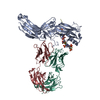
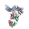
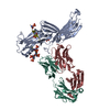
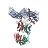
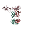
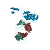
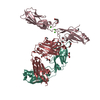
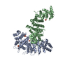
| Entry | Database: PDB / ID: 7dfb | ||||||
|---|---|---|---|---|---|---|---|
| Title | Crystal of Arrestin2-V2Rpp-6-7-Fab30 complex | ||||||
 Components Components |
| ||||||
 Keywords Keywords | SIGNALING PROTEIN / Arrestin / G-protein-coupled receptor / Phosphopeptide / Antibody fragment | ||||||
| Function / homology |  Function and homology information Function and homology informationTGFBR3 regulates TGF-beta signaling / MAP2K and MAPK activation / Activation of SMO / Golgi Associated Vesicle Biogenesis / Lysosome Vesicle Biogenesis / AP-2 adaptor complex binding / Ub-specific processing proteases / clathrin coat of coated pit / Cargo recognition for clathrin-mediated endocytosis / clathrin heavy chain binding ...TGFBR3 regulates TGF-beta signaling / MAP2K and MAPK activation / Activation of SMO / Golgi Associated Vesicle Biogenesis / Lysosome Vesicle Biogenesis / AP-2 adaptor complex binding / Ub-specific processing proteases / clathrin coat of coated pit / Cargo recognition for clathrin-mediated endocytosis / clathrin heavy chain binding / Clathrin-mediated endocytosis / clathrin-dependent endocytosis / desensitization of G protein-coupled receptor signaling pathway / acetylcholine receptor binding / G protein-coupled receptor internalization / inositol hexakisphosphate binding / sensory perception / Thrombin signalling through proteinase activated receptors (PARs) / G alpha (s) signalling events / clathrin binding / small molecule binding / phosphatidylinositol-3,4,5-trisphosphate binding / pseudopodium / positive regulation of receptor internalization / negative regulation of Notch signaling pathway / positive regulation of protein phosphorylation / receptor internalization / G protein-coupled receptor binding / protein transport / cytoplasmic vesicle / molecular adaptor activity / ubiquitin-dependent protein catabolic process / positive regulation of ERK1 and ERK2 cascade / signal transduction / nucleus / plasma membrane / cytoplasm / cytosol Similarity search - Function | ||||||
| Biological species |  synthetic construct (others) | ||||||
| Method |  X-RAY DIFFRACTION / SYNCHROTRON / MOLECULAR REPLACEMENT / Resolution: 3.28 Å X-RAY DIFFRACTION / SYNCHROTRON / MOLECULAR REPLACEMENT / Resolution: 3.28 Å | ||||||
 Authors Authors | Sun, J.P. / Yu, X. / Xiao, P. / He, Q.T. / Lin, J.Y. / Zhu, Z.L. | ||||||
| Funding support |  China, 1items China, 1items
| ||||||
 Citation Citation | Journal: Nat Commun / Year: 2021 Title: Structural studies of phosphorylation-dependent interactions between the V2R receptor and arrestin-2. Authors: He, Q.T. / Xiao, P. / Huang, S.M. / Jia, Y.L. / Zhu, Z.L. / Lin, J.Y. / Yang, F. / Tao, X.N. / Zhao, R.J. / Gao, F.Y. / Niu, X.G. / Xiao, K.H. / Wang, J. / Jin, C. / Sun, J.P. / Yu, X. | ||||||
| History |
|
- Structure visualization
| Structure viewer | Molecule: MolmilJmol/JSmol |
|---|
- Downloads & links
-Download
| PDBx/mmCIF format | 7dfb.cif.gz | 179.5 KB | Display | PDBx/mmCIF format |
|---|---|---|---|---|
| PDB format | pdb7dfb.ent.gz | 125.2 KB | Display | PDB format |
| PDBx/mmJSON format | 7dfb.json.gz | Tree view | PDBx/mmJSON format | |
| Others |  Other downloads Other downloads |
-Validation report
| Arichive directory | https://data.pdbj.org/pub/pdb/validation_reports/df/7dfbftp://data.pdbj.org/pub/pdb/validation_reports/df/7dfb | HTTPS FTP |
|---|
-Related structure data
| Related structure data |  7df9C  7dfaC  7dfcC 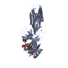 4jqiS S: Starting model for refinement C: citing same article ( |
|---|---|
| Similar structure data |
-Links
 PDBj
PDBj


- Assembly
| Deposited unit | 
| ||||||||||||
|---|---|---|---|---|---|---|---|---|---|---|---|---|---|
| 1 |
| ||||||||||||
| Unit cell |
|
-Components
| #1: Protein | Mass: 48272.832 Da / Num. of mol.: 1 Source method: isolated from a genetically manipulated source Source: (gene. exp.) Production host:  References: UniProt: P17870 |
|---|---|
| #2: Protein/peptide | Mass: 2916.511 Da / Num. of mol.: 1 / Source method: obtained synthetically / Source: (synth.) synthetic construct (others) |
| #3: Antibody | Mass: 24751.596 Da / Num. of mol.: 1 Source method: isolated from a genetically manipulated source Source: (gene. exp.) Production host: |
| #4: Antibody | Mass: 26828.885 Da / Num. of mol.: 1 Source method: isolated from a genetically manipulated source Source: (gene. exp.) Production host: |
| Has ligand of interest | Y |
| Has protein modification | Y |
-Experimental details
-Experiment
| Experiment | Method: X-RAY DIFFRACTION / Number of used crystals: 1 |
|---|
- Sample preparation
| Crystal | Density Matthews: 2.37 Å3/Da / Density % sol: 48.12 % |
|---|---|
| Crystal grow | Temperature: 289 K / Method: evaporation Details: 0% PEG 3350, 0.1M Succinic acid, pH 6.5-7.5, 0.2mM DMSO |
-Data collection
| Diffraction | Mean temperature: 100 K / Serial crystal experiment: N |
|---|---|
| Diffraction source | Source: SYNCHROTRON / Site: SSRF / Beamline: BL17B1 / Wavelength: 0.9776 Å |
| Detector | Type: DECTRIS PILATUS3 S 6M / Detector: PIXEL / Date: Nov 18, 2017 |
| Radiation | Protocol: SINGLE WAVELENGTH / Monochromatic (M) / Laue (L): M / Scattering type: x-ray |
| Radiation wavelength | Wavelength: 0.9776 Å / Relative weight: 1 |
| Reflection | Resolution: 3.28→45.36 Å / Num. obs: 30318 / % possible obs: 99.5 % / Redundancy: 6.5 % / Biso Wilson estimate: 111.16 Å2 / CC1/2: 0.999 / Rmerge(I) obs: 0.094 / Rpim(I) all: 0.04 / Net I/σ(I): 14 |
| Reflection shell | Resolution: 3.28→3.54 Å / Rmerge(I) obs: 0.929 / Num. unique obs: 3074 / CC1/2: 0.891 / Rpim(I) all: 0.376 |
- Processing
| Software |
| |||||||||||||||||||||||||||||||||||||||||||||||||||||||||||||||||||||||||||||
|---|---|---|---|---|---|---|---|---|---|---|---|---|---|---|---|---|---|---|---|---|---|---|---|---|---|---|---|---|---|---|---|---|---|---|---|---|---|---|---|---|---|---|---|---|---|---|---|---|---|---|---|---|---|---|---|---|---|---|---|---|---|---|---|---|---|---|---|---|---|---|---|---|---|---|---|---|---|---|
| Refinement | Method to determine structure: MOLECULAR REPLACEMENT Starting model: 4JQI Resolution: 3.28→45.36 Å / SU ML: 0.6236 / Cross valid method: FREE R-VALUE / σ(F): 0.25 / Phase error: 36.8775 / Stereochemistry target values: CDL v1.2
| |||||||||||||||||||||||||||||||||||||||||||||||||||||||||||||||||||||||||||||
| Solvent computation | Shrinkage radii: 0.9 Å / VDW probe radii: 1.11 Å / Solvent model: FLAT BULK SOLVENT MODEL | |||||||||||||||||||||||||||||||||||||||||||||||||||||||||||||||||||||||||||||
| Displacement parameters | Biso mean: 113.16 Å2 | |||||||||||||||||||||||||||||||||||||||||||||||||||||||||||||||||||||||||||||
| Refinement step | Cycle: LAST / Resolution: 3.28→45.36 Å
| |||||||||||||||||||||||||||||||||||||||||||||||||||||||||||||||||||||||||||||
| Refine LS restraints |
| |||||||||||||||||||||||||||||||||||||||||||||||||||||||||||||||||||||||||||||
| LS refinement shell |
|
