 Movie
Movie Controller


+ Open data

- Basic information
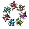
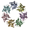
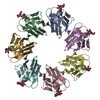
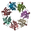
| Entry | Database: PDB / ID: 7ded | ||||||
|---|---|---|---|---|---|---|---|
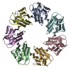
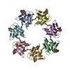
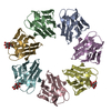
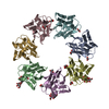
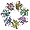
| Title | Mevo lectin complex with mannoheptose (Man7) | ||||||
 Components Components | lectin | ||||||
 Keywords Keywords | SUGAR BINDING PROTEIN / beta-prism I fold / Mevo lectin / heptamer / ring shape structure | ||||||
| Function / homology | Jacalin-like lectin domain / Aligned Prism / Vitelline Membrane Outer Layer Protein I, subunit A / Jacalin-like lectin domain / Jacalin-like lectin domain / Jacalin-like lectin domain superfamily / Mainly Beta / alpha-D-mannopyranose / Jacalin-type lectin domain-containing protein Function and homology information Function and homology information | ||||||
| Biological species |  Methanococcus voltae (archaea) Methanococcus voltae (archaea) | ||||||
| Method |  X-RAY DIFFRACTION / MOLECULAR REPLACEMENT / Resolution: 2.228 Å X-RAY DIFFRACTION / MOLECULAR REPLACEMENT / Resolution: 2.228 Å | ||||||
 Authors Authors | Sivaji, N. / Surolia, A. / Vijayan, M. | ||||||
| Funding support |  India, 1items India, 1items
| ||||||
 Citation Citation | Journal: Glycobiology / Year: 2021 Title: Mevo lectin specificity toward high-mannose structures with terminal alpha Man(1,2) alpha Man residues and its implication to inhibition of the entry of Mycobacterium tuberculosis into macrophages. Authors: Sivaji, N. / Harish, N. / Singh, S. / Singh, A. / Vijayan, M. / Surolia, A. #1: Journal: Glycobiology / Year: 2020Title: Structural and related studies on Mevo lectin from Methanococcus voltae A3. The first thorough characterisation of an archeal lectin and its interactions. Authors: Sivaji, N. / Suguna, K. / Surolia, A. / Vijayan, M. #2: Journal: Proteins / Year: 2016 Title: Archeal lectins: An identification through a genomic search. Authors: Abhinav, K.V. / Samuel, E. / Vijayan, M. | ||||||
| History |
|
- Structure visualization
| Structure viewer | Molecule: MolmilJmol/JSmol |
|---|
- Downloads & links
-Download
| PDBx/mmCIF format | 7ded.cif.gz | 199.7 KB | Display | PDBx/mmCIF format |
|---|---|---|---|---|
| PDB format | pdb7ded.ent.gz | 161.1 KB | Display | PDB format |
| PDBx/mmJSON format | 7ded.json.gz | Tree view | PDBx/mmJSON format | |
| Others |  Other downloads Other downloads |
-Validation report
| Arichive directory | https://data.pdbj.org/pub/pdb/validation_reports/de/7dedftp://data.pdbj.org/pub/pdb/validation_reports/de/7ded | HTTPS FTP |
|---|
-Related structure data
| Related structure data |  7bsbS S: Starting model for refinement |
|---|---|
| Similar structure data |


-Links
 PDBj
PDBj


- Assembly
| Deposited unit | 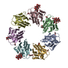
| ||||||||
|---|---|---|---|---|---|---|---|---|---|
| 1 |
| ||||||||
| Unit cell |
|
-Components
| #1: Protein | Mass: 15210.124 Da / Num. of mol.: 7 Source method: isolated from a genetically manipulated source Source: (gene. exp.) Methanococcus voltae (strain ATCC BAA-1334 / A3) (archaea)Strain: ATCC BAA-1334 / A3 / Gene: Mvol_0737 / Production host:  #2: Polysaccharide | #3: Sugar | ChemComp-MAN / |   Type: D-saccharide, alpha linking / Mass: 180.156 Da / Num. of mol.: 1 / Source method: obtained synthetically / Formula: C6H12O6 / Feature type: SUBJECT OF INVESTIGATION Type: D-saccharide, alpha linking / Mass: 180.156 Da / Num. of mol.: 1 / Source method: obtained synthetically / Formula: C6H12O6 / Feature type: SUBJECT OF INVESTIGATION#4: Chemical | ChemComp-EDO /   Mass: 62.068 Da / Num. of mol.: 15 / Source method: obtained synthetically / Formula: C2H6O2 Mass: 62.068 Da / Num. of mol.: 15 / Source method: obtained synthetically / Formula: C2H6O2#5: Water | ChemComp-HOH / |  Mass: 18.015 Da / Num. of mol.: 127 / Source method: isolated from a natural source / Formula: H2O Mass: 18.015 Da / Num. of mol.: 127 / Source method: isolated from a natural source / Formula: H2OHas ligand of interest | Y | |
|---|
-Experimental details
-Experiment
| Experiment | Method: X-RAY DIFFRACTION / Number of used crystals: 1 |
|---|
- Sample preparation
| Crystal | Density Matthews: 3.49 Å3/Da / Density % sol: 59.5 % |
|---|---|
| Crystal grow | Temperature: 295 K / Method: microbatch / pH: 6 Details: Tacsimate pH 6.0, 0.1 M MES monohydrate pH 6.0, 25 % (w/v) PEG 4000 |
-Data collection
| Diffraction | Mean temperature: 100 K / Serial crystal experiment: N |
|---|---|
| Diffraction source | Source: ROTATING ANODE / Type: BRUKER AXS MICROSTAR / Wavelength: 1.54 Å |
| Detector | Type: MAR scanner 345 mm plate / Detector: IMAGE PLATE / Date: Apr 14, 2020 |
| Radiation | Monochromator: Cu K / Protocol: SINGLE WAVELENGTH / Monochromatic (M) / Laue (L): M / Scattering type: x-ray |
| Radiation wavelength | Wavelength: 1.54 Å / Relative weight: 1 |
| Reflection | Resolution: 2.22→61.18 Å / Num. obs: 73523 / % possible obs: 99.7 % / Redundancy: 19.2 % / Biso Wilson estimate: 16.6 Å2 / CC1/2: 0.985 / Rmerge(I) obs: 0.39 / Rpim(I) all: 0.091 / Net I/σ(I): 7.8 |
| Reflection shell | Resolution: 2.23→2.35 Å / Redundancy: 17.6 % / Rmerge(I) obs: 0.813 / Mean I/σ(I) obs: 3.2 / Num. unique obs: 10366 / CC1/2: 0.524 / Rpim(I) all: 0.196 / % possible all: 98 |
- Processing
| Software |
| ||||||||||||||||||||||||||||||||||||||||||||||||||||||||||||||||||||||||||||||||||||||||||||||||||||||||||||||||||||||||||||||||||||||||||||||||||||||||||||||||
|---|---|---|---|---|---|---|---|---|---|---|---|---|---|---|---|---|---|---|---|---|---|---|---|---|---|---|---|---|---|---|---|---|---|---|---|---|---|---|---|---|---|---|---|---|---|---|---|---|---|---|---|---|---|---|---|---|---|---|---|---|---|---|---|---|---|---|---|---|---|---|---|---|---|---|---|---|---|---|---|---|---|---|---|---|---|---|---|---|---|---|---|---|---|---|---|---|---|---|---|---|---|---|---|---|---|---|---|---|---|---|---|---|---|---|---|---|---|---|---|---|---|---|---|---|---|---|---|---|---|---|---|---|---|---|---|---|---|---|---|---|---|---|---|---|---|---|---|---|---|---|---|---|---|---|---|---|---|---|---|---|---|
| Refinement | Method to determine structure: MOLECULAR REPLACEMENT Starting model: 7BSB Resolution: 2.228→59.592 Å / Cor.coef. Fo:Fc: 0.897 / Cor.coef. Fo:Fc free: 0.885 / WRfactor Rfree: 0.238 / WRfactor Rwork: 0.217 / Average fsc free: 0.8348 / Average fsc work: 0.841 / Cross valid method: THROUGHOUT / ESU R: 0.252 / ESU R Free: 0.203 Details: Hydrogens have been added in their riding positions
| ||||||||||||||||||||||||||||||||||||||||||||||||||||||||||||||||||||||||||||||||||||||||||||||||||||||||||||||||||||||||||||||||||||||||||||||||||||||||||||||||
| Solvent computation | Ion probe radii: 0.8 Å / Shrinkage radii: 0.8 Å / VDW probe radii: 1.2 Å / Solvent model: MASK BULK SOLVENT | ||||||||||||||||||||||||||||||||||||||||||||||||||||||||||||||||||||||||||||||||||||||||||||||||||||||||||||||||||||||||||||||||||||||||||||||||||||||||||||||||
| Displacement parameters | Biso mean: 19.837 Å2
| ||||||||||||||||||||||||||||||||||||||||||||||||||||||||||||||||||||||||||||||||||||||||||||||||||||||||||||||||||||||||||||||||||||||||||||||||||||||||||||||||
| Refinement step | Cycle: LAST / Resolution: 2.228→59.592 Å
| ||||||||||||||||||||||||||||||||||||||||||||||||||||||||||||||||||||||||||||||||||||||||||||||||||||||||||||||||||||||||||||||||||||||||||||||||||||||||||||||||
| Refine LS restraints |
| ||||||||||||||||||||||||||||||||||||||||||||||||||||||||||||||||||||||||||||||||||||||||||||||||||||||||||||||||||||||||||||||||||||||||||||||||||||||||||||||||
| LS refinement shell |
|
