 Movie
Movie Controller


+ Open data

- Basic information
| Entry | Database: PDB / ID: 7bsm | ||||||
|---|---|---|---|---|---|---|---|
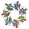
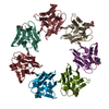
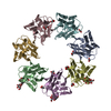
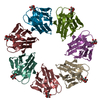
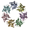
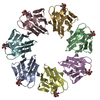
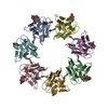
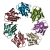
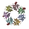
| Title | Mevo lectin complex with 2alpha-mannobiose | ||||||
 Components Components | lectin | ||||||
 Keywords Keywords | SUGAR BINDING PROTEIN / beta-prism I fold lectin / Archeal lectin / heptamer / ring shape structure | ||||||
| Function / homology |  Function and homology information Function and homology informationJacalin-like lectin domain / Aligned Prism / Vitelline Membrane Outer Layer Protein I, subunit A / Jacalin-like lectin domain / Jacalin-like lectin domain / Jacalin-like lectin domain superfamily / Mainly Beta Similarity search - Domain/homology | ||||||
| Biological species |  Methanococcus voltae (archaea) Methanococcus voltae (archaea) | ||||||
| Method |  X-RAY DIFFRACTION / SYNCHROTRON / MOLECULAR REPLACEMENT / Resolution: 2.8 Å X-RAY DIFFRACTION / SYNCHROTRON / MOLECULAR REPLACEMENT / Resolution: 2.8 Å | ||||||
 Authors Authors | Sivaji, N. / Suguna, K. / Surolia, A. / Vijayan, M. | ||||||
| Funding support |  India, 1items India, 1items
| ||||||
 Citation Citation | Journal: Glycobiology / Year: 2021 Title: Structural and related studies on Mevo lectin from Methanococcus voltae A3: the first thorough characterization of an archeal lectin and its interactions. Authors: Sivaji, N. / Suguna, K. / Surolia, A. / Vijayan, M. | ||||||
| History |
|
- Structure visualization
| Structure viewer | Molecule: MolmilJmol/JSmol |
|---|
- Downloads & links
-Download
| PDBx/mmCIF format | 7bsm.cif.gz | 371.8 KB | Display | PDBx/mmCIF format |
|---|---|---|---|---|
| PDB format | pdb7bsm.ent.gz | 302.5 KB | Display | PDB format |
| PDBx/mmJSON format | 7bsm.json.gz | Tree view | PDBx/mmJSON format | |
| Others |  Other downloads Other downloads |
-Validation report
| Arichive directory | https://data.pdbj.org/pub/pdb/validation_reports/bs/7bsmftp://data.pdbj.org/pub/pdb/validation_reports/bs/7bsm | HTTPS FTP |
|---|
-Related structure data
| Related structure data |  7bsbSC  7bsnC  7bt8C  7bt9C 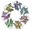 7bthC  7btlC S: Starting model for refinement C: citing same article ( |
|---|---|
| Similar structure data |

-Links
 PDBj
PDBj


- Assembly
| Deposited unit | 
| ||||||||
|---|---|---|---|---|---|---|---|---|---|
| 1 | 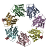
| ||||||||
| 2 | 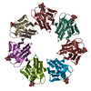
| ||||||||
| Unit cell |
|
-Components
| #1: Protein | Mass: 15783.812 Da / Num. of mol.: 14 Source method: isolated from a genetically manipulated source Source: (gene. exp.) Methanococcus voltae (strain ATCC BAA-1334 / A3) (archaea)Strain: ATCC BAA-1334 / A3 / Gene: Mvol_0737 / Production host:  #2: Polysaccharide | alpha-D-mannopyranose-(1-2)-alpha-D-mannopyranose / 2alpha-alpha-mannobiose   Source method: isolated from a genetically manipulated source Details: oligosaccharide / References: 2alpha-alpha-mannobiose #3: Sugar |   Type: D-saccharide, alpha linking / Mass: 180.156 Da / Num. of mol.: 3 Type: D-saccharide, alpha linking / Mass: 180.156 Da / Num. of mol.: 3Source method: isolated from a genetically manipulated source Formula: C6H12O6 #4: Chemical | ChemComp-ALA / |   Type: L-peptide linking / Mass: 89.093 Da / Num. of mol.: 1 / Source method: obtained synthetically / Formula: C3H7NO2 Type: L-peptide linking / Mass: 89.093 Da / Num. of mol.: 1 / Source method: obtained synthetically / Formula: C3H7NO2Has ligand of interest | Y | |
|---|
-Experimental details
-Experiment
| Experiment | Method: X-RAY DIFFRACTION / Number of used crystals: 1 |
|---|
- Sample preparation
| Crystal | Density Matthews: 2.85 Å3/Da / Density % sol: 56.9 % |
|---|---|
| Crystal grow | Temperature: 296 K / Method: microbatch Details: 6%(v/v) Tacsimate pH 6.0, 0.1 M MES monohydrate pH 6.0, 25%(w/v) polyethylene glycol 4000 PH range: 5.8-7.8 |
-Data collection
| Diffraction | Mean temperature: 100 K / Serial crystal experiment: N |
|---|---|
| Diffraction source | Source: SYNCHROTRON / Site: ELETTRA  / Beamline: 11.2C / Wavelength: 0.9767 Å / Beamline: 11.2C / Wavelength: 0.9767 Å |
| Detector | Type: DECTRIS PILATUS3 6M / Detector: PIXEL / Date: Dec 11, 2018 |
| Radiation | Protocol: SINGLE WAVELENGTH / Monochromatic (M) / Laue (L): M / Scattering type: x-ray |
| Radiation wavelength | Wavelength: 0.9767 Å / Relative weight: 1 |
| Reflection | Resolution: 2.8→96.25 Å / Num. obs: 70202 / % possible obs: 100 % / Redundancy: 13.2 % / CC1/2: 0.99 / Rmerge(I) obs: 0.22 / Net I/σ(I): 9 |
| Reflection shell | Resolution: 2.8→2.95 Å / Mean I/σ(I) obs: 1.8 / Num. unique obs: 10083 / CC1/2: 0.73 / % possible all: 100 |
- Processing
| Software |
| ||||||||||||||||||||||||||||||||||||||||||||||||||||||||||||||||||||||||||||||||||||||||||||||||||||||||||||||||||||||||||||||||||||||||||||||||||||||||||||||||||||||||||||||||||||||
|---|---|---|---|---|---|---|---|---|---|---|---|---|---|---|---|---|---|---|---|---|---|---|---|---|---|---|---|---|---|---|---|---|---|---|---|---|---|---|---|---|---|---|---|---|---|---|---|---|---|---|---|---|---|---|---|---|---|---|---|---|---|---|---|---|---|---|---|---|---|---|---|---|---|---|---|---|---|---|---|---|---|---|---|---|---|---|---|---|---|---|---|---|---|---|---|---|---|---|---|---|---|---|---|---|---|---|---|---|---|---|---|---|---|---|---|---|---|---|---|---|---|---|---|---|---|---|---|---|---|---|---|---|---|---|---|---|---|---|---|---|---|---|---|---|---|---|---|---|---|---|---|---|---|---|---|---|---|---|---|---|---|---|---|---|---|---|---|---|---|---|---|---|---|---|---|---|---|---|---|---|---|---|---|
| Refinement | Method to determine structure: MOLECULAR REPLACEMENT Starting model: 7BSB Resolution: 2.8→96.25 Å / Cor.coef. Fo:Fc: 0.929 / Cor.coef. Fo:Fc free: 0.92 / Cross valid method: THROUGHOUT / ESU R: 0.929 / ESU R Free: 0.35 Details: Hydrogens have been added in their riding positions
| ||||||||||||||||||||||||||||||||||||||||||||||||||||||||||||||||||||||||||||||||||||||||||||||||||||||||||||||||||||||||||||||||||||||||||||||||||||||||||||||||||||||||||||||||||||||
| Solvent computation | Ion probe radii: 0.8 Å / Shrinkage radii: 0.8 Å / VDW probe radii: 1.2 Å | ||||||||||||||||||||||||||||||||||||||||||||||||||||||||||||||||||||||||||||||||||||||||||||||||||||||||||||||||||||||||||||||||||||||||||||||||||||||||||||||||||||||||||||||||||||||
| Displacement parameters | Biso mean: 53.106 Å2
| ||||||||||||||||||||||||||||||||||||||||||||||||||||||||||||||||||||||||||||||||||||||||||||||||||||||||||||||||||||||||||||||||||||||||||||||||||||||||||||||||||||||||||||||||||||||
| Refinement step | Cycle: LAST / Resolution: 2.8→96.25 Å
| ||||||||||||||||||||||||||||||||||||||||||||||||||||||||||||||||||||||||||||||||||||||||||||||||||||||||||||||||||||||||||||||||||||||||||||||||||||||||||||||||||||||||||||||||||||||
| Refine LS restraints |
|