 Movie
Movie Controller

[English] 日本語
 Yorodumi
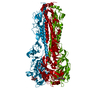
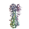
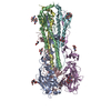
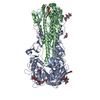
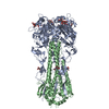
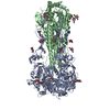
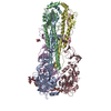
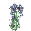
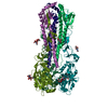
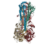
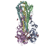
- PDB-7dea: Structure of an avian influenza H5 hemagglutinin from the influen... -
+ Open data

- Basic information
| Entry | Database: PDB / ID: 7dea | ||||||
|---|---|---|---|---|---|---|---|
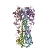
| Title | Structure of an avian influenza H5 hemagglutinin from the influenza virus A/duck Northern China/22/2017 (H5N6) | ||||||
 Components Components | (Hemagglutinin) x 2 | ||||||
 Keywords Keywords | VIRAL PROTEIN / H5N6 avian influenza virus / Haemagglutinin / Neuraminidase / Acid stability / Human infection | ||||||
| Function / homology |  Function and homology information Function and homology informationviral budding from plasma membrane / clathrin-dependent endocytosis of virus by host cell / apical plasma membrane / host cell surface receptor binding / fusion of virus membrane with host plasma membrane / fusion of virus membrane with host endosome membrane / viral envelope / virion attachment to host cell / host cell plasma membrane / virion membrane Similarity search - Function | ||||||
| Biological species |   Influenza A virus Influenza A virus | ||||||
| Method |  X-RAY DIFFRACTION / SYNCHROTRON / MOLECULAR REPLACEMENT / Resolution: 2.84 Å X-RAY DIFFRACTION / SYNCHROTRON / MOLECULAR REPLACEMENT / Resolution: 2.84 Å | ||||||
 Authors Authors | Sun, H. / Sun, H. / Song, J. / Zhang, W. / Wei, X. / Qi, J. / Gao, G.F. / Liu, J. | ||||||
 Citation Citation | Journal: To Be Published Title: Haemagglutinin and neuraminidase acid stability in H5N6 avian influenza virus confers infection adaptation in mammals Authors: Sun, H. / Sun, H. / Song, J. / Zhang, W. / Qi, J. / Gao, G.F. / Liu, J. | ||||||
| History |
|
- Structure visualization
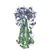
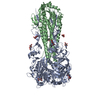
| Structure viewer | Molecule: MolmilJmol/JSmol |
|---|
- Downloads & links
-Download
| PDBx/mmCIF format | 7dea.cif.gz | 299.8 KB | Display | PDBx/mmCIF format |
|---|---|---|---|---|
| PDB format | pdb7dea.ent.gz | 245.8 KB | Display | PDB format |
| PDBx/mmJSON format | 7dea.json.gz | Tree view | PDBx/mmJSON format | |
| Others |  Other downloads Other downloads |
-Validation report
| Arichive directory | https://data.pdbj.org/pub/pdb/validation_reports/de/7deaftp://data.pdbj.org/pub/pdb/validation_reports/de/7dea | HTTPS FTP |
|---|
-Related structure data
| Related structure data |  7debC 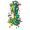 6ntfS S: Starting model for refinement C: citing same article ( |
|---|---|
| Similar structure data |

-Links
 PDBj
PDBj


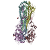
- Assembly
| Deposited unit | 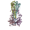
| ||||||||
|---|---|---|---|---|---|---|---|---|---|
| 1 |
| ||||||||
| Unit cell |
|
-Components
| #1: Protein | Mass: 36169.898 Da / Num. of mol.: 3 Source method: isolated from a genetically manipulated source Source: (gene. exp.) Influenza A virus / Gene: HAProduction host: Insect cell expression vector pTIE1 (others) #2: Protein | Mass: 19883.906 Da / Num. of mol.: 3 Source method: isolated from a genetically manipulated source Source: (gene. exp.) Influenza A virus / Gene: HAProduction host: Insect cell expression vector pTIE1 (others) References: UniProt: A0A6M2RI35 #3: Polysaccharide | Source method: isolated from a genetically manipulated source #4: Sugar | ChemComp-NAG /   Type: D-saccharide, beta linking / Mass: 221.208 Da / Num. of mol.: 6 Type: D-saccharide, beta linking / Mass: 221.208 Da / Num. of mol.: 6Source method: isolated from a genetically manipulated source Formula: C8H15NO6 / Feature type: SUBJECT OF INVESTIGATION #5: Water | ChemComp-HOH / |  Mass: 18.015 Da / Num. of mol.: 13 / Source method: isolated from a natural source / Formula: H2O Mass: 18.015 Da / Num. of mol.: 13 / Source method: isolated from a natural source / Formula: H2OHas ligand of interest | Y | Has protein modification | Y | |
|---|
-Experimental details
-Experiment
| Experiment | Method: X-RAY DIFFRACTION / Number of used crystals: 1 |
|---|
- Sample preparation
| Crystal | Density Matthews: 4.08 Å3/Da / Density % sol: 69.83 % |
|---|---|
| Crystal grow | Temperature: 291 K / Method: vapor diffusion, sitting drop Details: 0.1 M TRIS hydrochloride pH 8.5, 2.0 M Ammonium phosphate monobasic |
-Data collection
| Diffraction | Mean temperature: 100 K / Serial crystal experiment: N |
|---|---|
| Diffraction source | Source: SYNCHROTRON / Site: SSRF  / Beamline: BL17U1 / Wavelength: 0.97915 Å / Beamline: BL17U1 / Wavelength: 0.97915 Å |
| Detector | Type: ADSC QUANTUM 315r / Detector: CCD / Date: Jun 16, 2020 |
| Radiation | Protocol: SINGLE WAVELENGTH / Monochromatic (M) / Laue (L): M / Scattering type: x-ray |
| Radiation wavelength | Wavelength: 0.97915 Å / Relative weight: 1 |
| Reflection | Resolution: 2.7→135.91 Å / Num. obs: 65192 / % possible obs: 99.66 % / Redundancy: 6.8 % / Rmerge(I) obs: 0.166 / Net I/σ(I): 8.4 |
| Reflection shell | Resolution: 2.7→2.85 Å / Rmerge(I) obs: 0.785 / Mean I/σ(I) obs: 2.1 / Num. unique obs: 3762 |
- Processing
| Software |
| ||||||||||||||||||||
|---|---|---|---|---|---|---|---|---|---|---|---|---|---|---|---|---|---|---|---|---|---|
| Refinement | Method to determine structure: MOLECULAR REPLACEMENT Starting model: 6NTF Resolution: 2.84→135.91 Å / Cor.coef. Fo:Fc: 0.942 / Cor.coef. Fo:Fc free: 0.919 / SU B: 0.003 / SU ML: 0 / Cross valid method: THROUGHOUT / ESU R: 0.261 / ESU R Free: 0.309 / Stereochemistry target values: MAXIMUM LIKELIHOOD / Details: HYDROGENS HAVE BEEN ADDED IN THE RIDING POSITIONS
| ||||||||||||||||||||
| Solvent computation | Ion probe radii: 0.8 Å / Shrinkage radii: 0.8 Å / VDW probe radii: 1.2 Å / Solvent model: MASK | ||||||||||||||||||||
| Displacement parameters | Biso mean: 70.243 Å2
| ||||||||||||||||||||
| Refinement step | Cycle: 1 / Resolution: 2.84→135.91 Å
| ||||||||||||||||||||
| LS refinement shell | Resolution: 2.843→2.917 Å / Total num. of bins used: 20
|
