- PDB-7de8: Crystal Structure of outer membrane protein PorB with G103K mutat... -
+
Open data
ID or keywords:
Loading...
-
Basic information
Entry
Database: PDB / ID: 7de8
Title
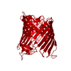
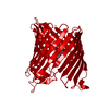
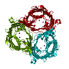
Crystal Structure of outer membrane protein PorB with G103K mutations from Neisseria meningitidis W135
Components
Outer membrane protein
Keywords
MEMBRANE PROTEIN / porin / outer membrane protein / membrane transport
Function / homology
Function and homology information
porin activity / pore complex / cell outer membrane / monoatomic ion transmembrane transport Similarity search - Function
Porin, Neisseria sp. type / Porin, Gram-negative type, conserved site / General diffusion Gram-negative porins signature. / Porin domain, Gram-negative type / Gram-negative porin / Porin, Gram-negative type / : / Porin domain superfamily Similarity search - Domain/homology
In the structure databanks used in Yorodumi, some data are registered as the other names, "COVID-19 virus" and "2019-nCoV". Here are the details of the virus and the list of structure data.
Jan 31, 2019. EMDB accession codes are about to change! (news from PDBe EMDB page)
EMDB accession codes are about to change! (news from PDBe EMDB page)
The allocation of 4 digits for EMDB accession codes will soon come to an end. Whilst these codes will remain in use, new EMDB accession codes will include an additional digit and will expand incrementally as the available range of codes is exhausted. The current 4-digit format prefixed with “EMD-” (i.e. EMD-XXXX) will advance to a 5-digit format (i.e. EMD-XXXXX), and so on. It is currently estimated that the 4-digit codes will be depleted around Spring 2019, at which point the 5-digit format will come into force.
The EM Navigator/Yorodumi systems omit the EMD- prefix.
Related info.:Q: What is EMD? / ID/Accession-code notation in Yorodumi/EM Navigator
Yorodumi is a browser for structure data from EMDB, PDB, SASBDB, etc.
This page is also the successor to EM Navigator detail page, and also detail information page/front-end page for Omokage search.
The word "yorodu" (or yorozu) is an old Japanese word meaning "ten thousand". "mi" (miru) is to see.
Related info.:EMDB / PDB / SASBDB / Comparison of 3 databanks / Yorodumi Search / Aug 31, 2016. New EM Navigator & Yorodumi / Yorodumi Papers / Jmol/JSmol / Function and homology information / Changes in new EM Navigator and Yorodumi
 Movie
Movie Controller

 Yorodumi
 Open data
 Basic information
 Components
 Keywords
 Function and homology information
 Neisseria meningitidis (bacteria)
 X-RAY DIFFRACTION /
  Authors
 Germany,
  Japan, 2items
  Citation
 Structure visualization
 Downloads & links
 Other downloads


 PDBj
PDBj Assembly
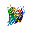

 Sample preparation
 / Beamline: X06SA / Wavelength: 1 Å
 Processing