 Movie
Movie Controller

[English] 日本語
 Yorodumi
- PDB-3vzw: Crystal Structure of outer membrane protein PorB from Neisseria m... -
+ Open data

- Basic information
| Entry | Database: PDB / ID: 3vzw | |||||||||
|---|---|---|---|---|---|---|---|---|---|---|
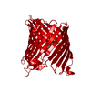
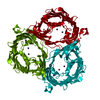
| Title | Crystal Structure of outer membrane protein PorB from Neisseria meningitidis in complex with galactose | |||||||||
 Components Components | outer membrane proteins | |||||||||
 Keywords Keywords | MEMBRANE PROTEIN / transport | |||||||||
| Function / homology |  Function and homology information Function and homology informationporin activity / pore complex / cell outer membrane / monoatomic ion transmembrane transport Similarity search - Function | |||||||||
| Biological species |  Neisseria meningitidis (bacteria) Neisseria meningitidis (bacteria) | |||||||||
| Method |  X-RAY DIFFRACTION / SYNCHROTRON / MOLECULAR REPLACEMENT / Resolution: 3.2 Å X-RAY DIFFRACTION / SYNCHROTRON / MOLECULAR REPLACEMENT / Resolution: 3.2 Å | |||||||||
 Authors Authors | Tanabe, M. / Iverson, T.M. | |||||||||
 Citation Citation | Journal: Proc.Natl.Acad.Sci.USA / Year: 2010 Title: Structural basis for solute transport, nucleotide regulation, and immunological recognition of Neisseria meningitidis PorB. Authors: Tanabe, M. / Nimigean, C.M. / Iverson, T.M. | |||||||||
| History |
|
- Structure visualization
| Structure viewer | Molecule: MolmilJmol/JSmol |
|---|
- Downloads & links
-Download
| PDBx/mmCIF format | 3vzw.cif.gz | 79.7 KB | Display | PDBx/mmCIF format |
|---|---|---|---|---|
| PDB format | pdb3vzw.ent.gz | 58 KB | Display | PDB format |
| PDBx/mmJSON format | 3vzw.json.gz | Tree view | PDBx/mmJSON format | |
| Others |  Other downloads Other downloads |
-Validation report
| Arichive directory | https://data.pdbj.org/pub/pdb/validation_reports/vz/3vzwftp://data.pdbj.org/pub/pdb/validation_reports/vz/3vzw | HTTPS FTP |
|---|
-Related structure data
| Related structure data |  3a2sC  3vztC  3vzuC  3a2r C: citing same article ( S: Starting model for refinement |
|---|---|
| Similar structure data |


-Links
 PDBj
PDBj

- Assembly
| Deposited unit | 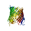
| ||||||||
|---|---|---|---|---|---|---|---|---|---|
| 1 | 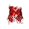
| ||||||||
| Unit cell |
|
-Components
| #1: Protein | Mass: 38179.305 Da / Num. of mol.: 1 Source method: isolated from a genetically manipulated source Source: (gene. exp.) Neisseria meningitidis (bacteria) / Strain: W135 / Production host: | ||
|---|---|---|---|
| #2: Sugar | ChemComp-GAL /   Type: D-saccharide, beta linking / Mass: 180.156 Da / Num. of mol.: 1 Type: D-saccharide, beta linking / Mass: 180.156 Da / Num. of mol.: 1Source method: isolated from a genetically manipulated source Formula: C6H12O6 | ||
| #3: Chemical |   Mass: 132.905 Da / Num. of mol.: 2 / Source method: obtained synthetically / Formula: Cs Mass: 132.905 Da / Num. of mol.: 2 / Source method: obtained synthetically / Formula: CsSequence details | A SEQUENCE REFERENCE FOR THIS PROTEIN DOES NOT CURRENTLY EXIST. | |
-Experimental details
-Experiment
| Experiment | Method: X-RAY DIFFRACTION / Number of used crystals: 1 |
|---|
- Sample preparation
| Crystal | Density Matthews: 2.85 Å3/Da / Density % sol: 56.83 % |
|---|---|
| Crystal grow | Temperature: 291 K / Method: vapor diffusion, hanging drop / pH: 6 Details: 100mM MES, 50mM CsCl, 28-32% Jeffamine M-600, pH6.0, VAPOR DIFFUSION, HANGING DROP, temperature 291K |
-Data collection
| Diffraction | Mean temperature: 100 K |
|---|---|
| Diffraction source | Source: SYNCHROTRON / Site: APS  / Beamline: 21-ID-D / Wavelength: 0.978 Å / Beamline: 21-ID-D / Wavelength: 0.978 Å |
| Detector | Type: MARMOSAIC 300 mm CCD / Detector: CCD / Date: Jan 31, 2009 |
| Radiation | Monochromator: Si(III) / Protocol: SINGLE WAVELENGTH / Monochromatic (M) / Laue (L): M / Scattering type: x-ray |
| Radiation wavelength | Wavelength: 0.978 Å / Relative weight: 1 |
| Reflection | Resolution: 3.2→50 Å / Num. all: 7136 / Num. obs: 6570 / % possible obs: 91.7 % / Observed criterion σ(F): 3 / Observed criterion σ(I): 3 |
| Reflection shell | Lowest resolution: 3.28 Å / Redundancy: 3.1 % / Mean I/σ(I) obs: 5.9 / Num. unique all: 264 / Rsym value: 0.148 / % possible all: 51.5 |
- Processing
| Software |
| ||||||||||||||||||||||||||||||||||||||||||||||||||||||||||||||||||||||||||||||||||||||||||||||||||||||||||||||||||||||||||||||||||||||||||||||||||||||||||||||||||||||||||
|---|---|---|---|---|---|---|---|---|---|---|---|---|---|---|---|---|---|---|---|---|---|---|---|---|---|---|---|---|---|---|---|---|---|---|---|---|---|---|---|---|---|---|---|---|---|---|---|---|---|---|---|---|---|---|---|---|---|---|---|---|---|---|---|---|---|---|---|---|---|---|---|---|---|---|---|---|---|---|---|---|---|---|---|---|---|---|---|---|---|---|---|---|---|---|---|---|---|---|---|---|---|---|---|---|---|---|---|---|---|---|---|---|---|---|---|---|---|---|---|---|---|---|---|---|---|---|---|---|---|---|---|---|---|---|---|---|---|---|---|---|---|---|---|---|---|---|---|---|---|---|---|---|---|---|---|---|---|---|---|---|---|---|---|---|---|---|---|---|---|---|---|
| Refinement | Method to determine structure: MOLECULAR REPLACEMENT Starting model: 3A2R 3a2r Resolution: 3.2→42.18 Å / Cor.coef. Fo:Fc: 0.935 / Cor.coef. Fo:Fc free: 0.937 / Cross valid method: THROUGHOUT / ESU R Free: 0.546 / Stereochemistry target values: MAXIMUM LIKELIHOOD / Details: HYDROGENS HAVE BEEN USED IF PRESENT IN THE INPUT
| ||||||||||||||||||||||||||||||||||||||||||||||||||||||||||||||||||||||||||||||||||||||||||||||||||||||||||||||||||||||||||||||||||||||||||||||||||||||||||||||||||||||||||
| Solvent computation | Ion probe radii: 0.8 Å / Shrinkage radii: 0.8 Å / VDW probe radii: 1.2 Å / Solvent model: MASK | ||||||||||||||||||||||||||||||||||||||||||||||||||||||||||||||||||||||||||||||||||||||||||||||||||||||||||||||||||||||||||||||||||||||||||||||||||||||||||||||||||||||||||
| Displacement parameters | Biso mean: 125.775 Å2
| ||||||||||||||||||||||||||||||||||||||||||||||||||||||||||||||||||||||||||||||||||||||||||||||||||||||||||||||||||||||||||||||||||||||||||||||||||||||||||||||||||||||||||
| Refinement step | Cycle: LAST / Resolution: 3.2→42.18 Å
| ||||||||||||||||||||||||||||||||||||||||||||||||||||||||||||||||||||||||||||||||||||||||||||||||||||||||||||||||||||||||||||||||||||||||||||||||||||||||||||||||||||||||||
| Refine LS restraints |
| ||||||||||||||||||||||||||||||||||||||||||||||||||||||||||||||||||||||||||||||||||||||||||||||||||||||||||||||||||||||||||||||||||||||||||||||||||||||||||||||||||||||||||
| LS refinement shell | Resolution: 3.2→3.283 Å / Total num. of bins used: 20
|
