 Movie
Movie Controller


+ Open data

- Basic information
| Entry | Database: PDB / ID: 7ddy | ||||||
|---|---|---|---|---|---|---|---|
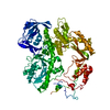
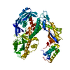
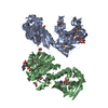
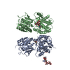
| Title | Crystal structure of an acetyl xylan esterase AlAXEase | ||||||
 Components Components | G-D-S-L family lipolytic protein | ||||||
 Keywords Keywords | HYDROLASE / Acetyl xylan esterase / SGNH / carbohydrate esterase | ||||||
| Function / homology | Lipase, GDSL, active site / Lipolytic enzymes "G-D-S-L" family, serine active site. / : / SGNH hydrolase-type esterase domain / GDSL-like Lipase/Acylhydrolase family / SGNH hydrolase superfamily / phosphatidylcholine lysophospholipase A1 activity / lipid metabolic process / G-D-S-L family lipolytic protein Function and homology information Function and homology information | ||||||
| Biological species |  Arcticibacterium luteifluviistationis (bacteria) Arcticibacterium luteifluviistationis (bacteria) | ||||||
| Method |  X-RAY DIFFRACTION / SYNCHROTRON / SAD / Resolution: 2.505 Å X-RAY DIFFRACTION / SYNCHROTRON / SAD / Resolution: 2.505 Å | ||||||
 Authors Authors | Zhang, Y. / Li, P.Y. / Zhang, Y.Z. | ||||||
 Citation Citation | Journal: J.Biol.Chem. / Year: 2021 Title: Active site architecture of an acetyl xylan esterase indicates a novel cold adaptation strategy. Authors: Zhang, Y. / Ding, H.T. / Jiang, W.X. / Zhang, X. / Cao, H.Y. / Wang, J.P. / Li, C.Y. / Huang, F. / Zhang, X.Y. / Chen, X.L. / Zhang, Y.Z. / Li, P.Y. | ||||||
| History |
|
- Structure visualization
| Structure viewer | Molecule: MolmilJmol/JSmol |
|---|
- Downloads & links
-Download
| PDBx/mmCIF format | 7ddy.cif.gz | 175.2 KB | Display | PDBx/mmCIF format |
|---|---|---|---|---|
| PDB format | pdb7ddy.ent.gz | 139 KB | Display | PDB format |
| PDBx/mmJSON format | 7ddy.json.gz | Tree view | PDBx/mmJSON format | |
| Others |  Other downloads Other downloads |
-Validation report
| Arichive directory | https://data.pdbj.org/pub/pdb/validation_reports/dd/7ddyftp://data.pdbj.org/pub/pdb/validation_reports/dd/7ddy | HTTPS FTP |
|---|
-Related structure data
| Similar structure data |
|---|


-Links
 PDBj
PDBj- Assembly
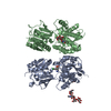
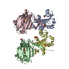
| Deposited unit | 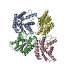
| ||||||||
|---|---|---|---|---|---|---|---|---|---|
| 1 |
| ||||||||
| Unit cell |
|
-Components
| #1: Protein | Mass: 26271.020 Da / Num. of mol.: 4 Source method: isolated from a genetically manipulated source Source: (gene. exp.) Arcticibacterium luteifluviistationis (bacteria)Gene: DJ013_06310 / Production host: #2: Water | ChemComp-HOH / |  Mass: 18.015 Da / Num. of mol.: 506 / Source method: isolated from a natural source / Formula: H2O Mass: 18.015 Da / Num. of mol.: 506 / Source method: isolated from a natural source / Formula: H2O |
|---|
-Experimental details
-Experiment
| Experiment | Method: X-RAY DIFFRACTION / Number of used crystals: 1 |
|---|
- Sample preparation
| Crystal | Density Matthews: 2.36 Å3/Da / Density % sol: 47.8 % |
|---|---|
| Crystal grow | Temperature: 291 K / Method: vapor diffusion, hanging drop / Details: 15% PEG 3350 0.1 M succinic acid |
-Data collection
| Diffraction | Mean temperature: 100 K / Serial crystal experiment: N |
|---|---|
| Diffraction source | Source: SYNCHROTRON / Site: SSRF  / Beamline: BL17U1 / Wavelength: 0.9791 Å / Beamline: BL17U1 / Wavelength: 0.9791 Å |
| Detector | Type: ADSC QUANTUM 315r / Detector: CCD / Date: Mar 21, 2017 |
| Radiation | Protocol: SINGLE WAVELENGTH / Monochromatic (M) / Laue (L): M / Scattering type: x-ray |
| Radiation wavelength | Wavelength: 0.9791 Å / Relative weight: 1 |
| Reflection | Resolution: 2.5→50 Å / Num. obs: 33141 / % possible obs: 98.8 % / Redundancy: 3.4 % / Rmerge(I) obs: 0.137 / Rpim(I) all: 0.086 / Rrim(I) all: 0.162 / Net I/σ(I): 8.25 |
| Reflection shell | Resolution: 2.5→2.54 Å / Rmerge(I) obs: 0.306 / Mean I/σ(I) obs: 2.67 / Num. unique obs: 3086 / Rpim(I) all: 0.192 / Rrim(I) all: 0.362 |
- Processing
| Software |
| ||||||||||||||||||||||||||||||||||||||||||||||||||||||||||||||||||||||||||||||
|---|---|---|---|---|---|---|---|---|---|---|---|---|---|---|---|---|---|---|---|---|---|---|---|---|---|---|---|---|---|---|---|---|---|---|---|---|---|---|---|---|---|---|---|---|---|---|---|---|---|---|---|---|---|---|---|---|---|---|---|---|---|---|---|---|---|---|---|---|---|---|---|---|---|---|---|---|---|---|---|
| Refinement | Method to determine structure: SAD / Resolution: 2.505→49.327 Å / SU ML: 0.25 / Cross valid method: THROUGHOUT / σ(F): 1.34 / Phase error: 23.15 / Stereochemistry target values: ML
| ||||||||||||||||||||||||||||||||||||||||||||||||||||||||||||||||||||||||||||||
| Solvent computation | Shrinkage radii: 0.9 Å / VDW probe radii: 1.11 Å / Solvent model: FLAT BULK SOLVENT MODEL | ||||||||||||||||||||||||||||||||||||||||||||||||||||||||||||||||||||||||||||||
| Displacement parameters | Biso max: 76.98 Å2 / Biso mean: 28.207 Å2 / Biso min: 11.29 Å2 | ||||||||||||||||||||||||||||||||||||||||||||||||||||||||||||||||||||||||||||||
| Refinement step | Cycle: final / Resolution: 2.505→49.327 Å
| ||||||||||||||||||||||||||||||||||||||||||||||||||||||||||||||||||||||||||||||
| Refine LS restraints |
| ||||||||||||||||||||||||||||||||||||||||||||||||||||||||||||||||||||||||||||||
| LS refinement shell | Refine-ID: X-RAY DIFFRACTION / Rfactor Rfree error: 0
|
