 Movie
Movie Controller

[English] 日本語
 Yorodumi
- PDB-7dda: Envelope protein VP37 a crystal structure from White Spot Syndrom... -
+ Open data

- Basic information
| Entry | Database: PDB / ID: 7dda | ||||||
|---|---|---|---|---|---|---|---|
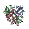
| Title | Envelope protein VP37 a crystal structure from White Spot Syndrome Virus | ||||||
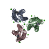
 Components Components | Envelope protein | ||||||
 Keywords Keywords | VIRAL PROTEIN / Envelope protein / Sulfate binding site / VP281 / VP37 / White spot syndrome virus (WSSV) | ||||||
| Function / homology | viral envelope / Envelope protein Function and homology information Function and homology information | ||||||
| Biological species |  White spot syndrome virus White spot syndrome virus | ||||||
| Method |  X-RAY DIFFRACTION / SYNCHROTRON / SAD / Resolution: 2.51 Å X-RAY DIFFRACTION / SYNCHROTRON / SAD / Resolution: 2.51 Å | ||||||
 Authors Authors | Somsoros, W. / Sangawa, T. / Takebe, K. / Attarataya, J. / Suzuki, M. / Khunrae, P. | ||||||
 Citation Citation | Journal: J.Gen.Virol. / Year: 2021 Title: Crystal structure of the C-terminal domain of envelope protein VP37 from white spot syndrome virus reveals sulphate binding sites responsible for heparin binding. Authors: Somsoros, W. / Sangawa, T. / Takebe, K. / Attarataya, J. / Wongprasert, K. / Senapin, S. / Rattanarojpong, T. / Suzuki, M. / Khunrae, P. | ||||||
| History |
|
- Structure visualization
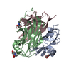
| Structure viewer | Molecule: MolmilJmol/JSmol |
|---|
- Downloads & links
-Download
| PDBx/mmCIF format | 7dda.cif.gz | 45.5 KB | Display | PDBx/mmCIF format |
|---|---|---|---|---|
| PDB format | pdb7dda.ent.gz | 25.1 KB | Display | PDB format |
| PDBx/mmJSON format | 7dda.json.gz | Tree view | PDBx/mmJSON format | |
| Others |  Other downloads Other downloads |
-Validation report
| Arichive directory | https://data.pdbj.org/pub/pdb/validation_reports/dd/7ddaftp://data.pdbj.org/pub/pdb/validation_reports/dd/7dda | HTTPS FTP |
|---|
-Related structure data
| Similar structure data |
|---|


-Links
 PDBj
PDBj- Assembly
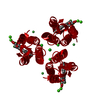
| Deposited unit | 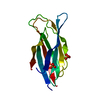
| ||||||||||||
|---|---|---|---|---|---|---|---|---|---|---|---|---|---|
| 1 | 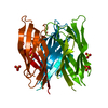
| ||||||||||||
| Unit cell |
| ||||||||||||
| Components on special symmetry positions |
|
-Components
| #1: Protein | Mass: 18984.098 Da / Num. of mol.: 1 Source method: isolated from a genetically manipulated source Source: (gene. exp.) White spot syndrome virus / Gene: VP281 / Plasmid: pET15bThioDetails (production host): pET15bThio, a pET15b (+) from Novagen which was modified to have thioredoxin and TEV cleavage site encoding sequence inserted upstream of the multiple cloning region Production host:  | ||||||
|---|---|---|---|---|---|---|---|
| #2: Chemical |   Mass: 96.063 Da / Num. of mol.: 2 / Source method: obtained synthetically / Formula: SO4 / Feature type: SUBJECT OF INVESTIGATION Mass: 96.063 Da / Num. of mol.: 2 / Source method: obtained synthetically / Formula: SO4 / Feature type: SUBJECT OF INVESTIGATION#3: Water | ChemComp-HOH / |  Mass: 18.015 Da / Num. of mol.: 13 / Source method: isolated from a natural source / Formula: H2O Mass: 18.015 Da / Num. of mol.: 13 / Source method: isolated from a natural source / Formula: H2OHas ligand of interest | Y | Has protein modification | Y | |
-Experimental details
-Experiment
| Experiment | Method: X-RAY DIFFRACTION / Number of used crystals: 1 |
|---|
- Sample preparation
| Crystal | Density Matthews: 3.01 Å3/Da / Density % sol: 59.11 % / Description: Hexagonal shaped crystals |
|---|---|
| Crystal grow | Temperature: 293.2 K / Method: vapor diffusion, hanging drop / pH: 6.5 Details: 1.5 M Ammonium sulfate, 100 mM Bis-Tris pH 6.5, 100 mM NaCl, VAPOR DIFFUSION, HANGING DROP, temperature 293.2K |
-Data collection
| Diffraction | Mean temperature: 100 K / Serial crystal experiment: N |
|---|---|
| Diffraction source | Source: SYNCHROTRON / Site: Photon Factory  / Beamline: BL-17A / Wavelength: 0.98 Å / Beamline: BL-17A / Wavelength: 0.98 Å |
| Detector | Type: DECTRIS PILATUS 6M / Detector: PIXEL / Date: Mar 9, 2016 |
| Radiation | Monochromator: Si(111) double crystal monochromator / Protocol: SINGLE WAVELENGTH / Monochromatic (M) / Laue (L): M / Scattering type: x-ray |
| Radiation wavelength | Wavelength: 0.98 Å / Relative weight: 1 |
| Reflection | Resolution: 2.505→44.691 Å / Num. obs: 8198 / % possible obs: 96.2 % / Redundancy: 37.1 % / Biso Wilson estimate: 28.51 Å2 / CC1/2: 1 / CC star: 1 / Rmerge(I) obs: 0.085 / Rpim(I) all: 0.014 / Rrim(I) all: 0.086 / Net I/σ(I): 43.6 |
| Reflection shell | Resolution: 2.505→2.548 Å / Redundancy: 36.1 % / Rmerge(I) obs: 0.327 / Mean I/σ(I) obs: 12.2 / Num. unique obs: 410 / CC1/2: 0.994 / CC star: 0.998 / Rpim(I) all: 0.054 / Rrim(I) all: 0.331 / % possible all: 100 |
- Processing
| Software |
| |||||||||||||||||||||||||||||||||||||||||||||||||||||||||||||||||||||||||||||
|---|---|---|---|---|---|---|---|---|---|---|---|---|---|---|---|---|---|---|---|---|---|---|---|---|---|---|---|---|---|---|---|---|---|---|---|---|---|---|---|---|---|---|---|---|---|---|---|---|---|---|---|---|---|---|---|---|---|---|---|---|---|---|---|---|---|---|---|---|---|---|---|---|---|---|---|---|---|---|
| Refinement | Method to determine structure: SAD / Resolution: 2.51→40.74 Å / SU ML: 0.2879 / Cross valid method: FREE R-VALUE / σ(F): 1.36 / Phase error: 23.876 Stereochemistry target values: GeoStd + Monomer Library + CDL v1.2
| |||||||||||||||||||||||||||||||||||||||||||||||||||||||||||||||||||||||||||||
| Solvent computation | Shrinkage radii: 0.9 Å / VDW probe radii: 1.11 Å / Solvent model: FLAT BULK SOLVENT MODEL | |||||||||||||||||||||||||||||||||||||||||||||||||||||||||||||||||||||||||||||
| Displacement parameters | Biso mean: 29.26 Å2 | |||||||||||||||||||||||||||||||||||||||||||||||||||||||||||||||||||||||||||||
| Refinement step | Cycle: LAST / Resolution: 2.51→40.74 Å
| |||||||||||||||||||||||||||||||||||||||||||||||||||||||||||||||||||||||||||||
| Refine LS restraints |
| |||||||||||||||||||||||||||||||||||||||||||||||||||||||||||||||||||||||||||||
| LS refinement shell |
|
