 Movie
Movie Controller


+ Open data

- Basic information
| Entry | Database: PDB / ID: 6wzc | |||||||||
|---|---|---|---|---|---|---|---|---|---|---|
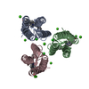
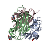
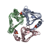
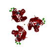
| Title | Ni-bound structure of an engineered protein trimer, TriCyt3 | |||||||||
 Components Components | Soluble cytochrome b562 | |||||||||
 Keywords Keywords | METAL BINDING PROTEIN / four-helix bundle / metalloprotein trimer | |||||||||
| Function / homology |  Function and homology information Function and homology informationelectron transport chain / periplasmic space / electron transfer activity / iron ion binding / heme binding Similarity search - Function | |||||||||
| Biological species |  | |||||||||
| Method |  X-RAY DIFFRACTION / SYNCHROTRON / MOLECULAR REPLACEMENT / Resolution: 2.195 Å X-RAY DIFFRACTION / SYNCHROTRON / MOLECULAR REPLACEMENT / Resolution: 2.195 Å | |||||||||
 Authors Authors | Tezcan, F.A. / Kakkis, A. | |||||||||
| Funding support |  United States, 2items United States, 2items
| |||||||||
 Citation Citation | Journal: Angew.Chem.Int.Ed.Engl. / Year: 2020 Title: Metal-Templated Design of Chemically Switchable Protein Assemblies with High-Affinity Coordination Sites. Authors: Kakkis, A. / Gagnon, D. / Esselborn, J. / Britt, R.D. / Tezcan, F.A. | |||||||||
| History |
|
- Structure visualization
| Structure viewer | Molecule: MolmilJmol/JSmol |
|---|
- Downloads & links
-Download
| PDBx/mmCIF format | 6wzc.cif.gz | 41.9 KB | Display | PDBx/mmCIF format |
|---|---|---|---|---|
| PDB format | pdb6wzc.ent.gz | 26.3 KB | Display | PDB format |
| PDBx/mmJSON format | 6wzc.json.gz | Tree view | PDBx/mmJSON format | |
| Others |  Other downloads Other downloads |
-Validation report
| Arichive directory | https://data.pdbj.org/pub/pdb/validation_reports/wz/6wzcftp://data.pdbj.org/pub/pdb/validation_reports/wz/6wzc | HTTPS FTP |
|---|
-Related structure data
| Related structure data | 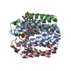 6wyuC 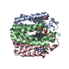 6wz0C  6wz1C 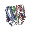 6wz2C 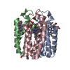 6wz3C  6wz7C 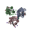 6wzaC 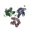 6x7eC  6x8xC 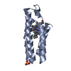 2bc5S S: Starting model for refinement C: citing same article ( |
|---|---|
| Similar structure data |


-Links
 PDBj
PDBj


- Assembly
| Deposited unit | 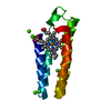
| |||||||||||||||||||||||||||
|---|---|---|---|---|---|---|---|---|---|---|---|---|---|---|---|---|---|---|---|---|---|---|---|---|---|---|---|---|
| 1 | 
| |||||||||||||||||||||||||||
| Unit cell |
| |||||||||||||||||||||||||||
| Components on special symmetry positions |
|
-Components
-Protein , 1 types, 1 molecules A
| #1: Protein | Mass: 11796.407 Da / Num. of mol.: 1 Mutation: T31K, A35K, Q41K, D54A, K59I, H63V, I67E, V69A, G70W, Q71E, D73H, L76A, K77H, N80K, R98C, Y101C Source method: isolated from a genetically manipulated source Source: (gene. exp.) |
|---|
-Non-polymers , 5 types, 115 molecules 


| #2: Chemical | ChemComp-HEC /  Mass: 618.503 Da / Num. of mol.: 1 / Source method: obtained synthetically / Formula: C34H34FeN4O4 Mass: 618.503 Da / Num. of mol.: 1 / Source method: obtained synthetically / Formula: C34H34FeN4O4 | ||||||
|---|---|---|---|---|---|---|---|
| #3: Chemical |  Mass: 58.693 Da / Num. of mol.: 2 / Source method: obtained synthetically / Formula: Ni / Feature type: SUBJECT OF INVESTIGATION Mass: 58.693 Da / Num. of mol.: 2 / Source method: obtained synthetically / Formula: Ni / Feature type: SUBJECT OF INVESTIGATION#4: Chemical | ChemComp-CA /  Mass: 40.078 Da / Num. of mol.: 4 / Source method: obtained synthetically / Formula: Ca Mass: 40.078 Da / Num. of mol.: 4 / Source method: obtained synthetically / Formula: Ca#5: Chemical |  Mass: 35.453 Da / Num. of mol.: 3 / Source method: obtained synthetically / Formula: Cl Mass: 35.453 Da / Num. of mol.: 3 / Source method: obtained synthetically / Formula: Cl#6: Water | ChemComp-HOH / | Mass: 18.015 Da / Num. of mol.: 105 / Source method: isolated from a natural source / Formula: H2O |
-Details
| Has ligand of interest | Y |
|---|---|
| Has protein modification | Y |
-Experimental details
-Experiment
| Experiment | Method: X-RAY DIFFRACTION / Number of used crystals: 1 |
|---|
- Sample preparation
| Crystal | Density Matthews: 4.34 Å3/Da / Density % sol: 71.66 % |
|---|---|
| Crystal grow | Temperature: 298 K / Method: vapor diffusion, sitting drop / pH: 6.5 / Details: 25% PEG2000, 0.2 M CaCl2, 0.1 M Bis-Tris (pH 6.5) |
-Data collection
| Diffraction | Mean temperature: 100 K / Serial crystal experiment: N |
|---|---|
| Diffraction source | Source: SYNCHROTRON / Site: SSRL / Beamline: BL12-2 / Wavelength: 1.2398 Å |
| Detector | Type: DECTRIS PILATUS 6M / Detector: PIXEL / Date: Mar 6, 2020 |
| Radiation | Protocol: SINGLE WAVELENGTH / Monochromatic (M) / Laue (L): M / Scattering type: x-ray |
| Radiation wavelength | Wavelength: 1.2398 Å / Relative weight: 1 |
| Reflection | Resolution: 2.195→35.7108 Å / Num. obs: 10934 / % possible obs: 99.62 % / Redundancy: 12.7 % / CC1/2: 0.887 / Rmerge(I) obs: 0.7438 / Rpim(I) all: 0.1867 / Rrim(I) all: 0.7678 / Net I/σ(I): 2.96 |
| Reflection shell | Resolution: 2.195→2.274 Å / Rmerge(I) obs: 0.5781 / Num. unique obs: 1068 / CC1/2: 0.887 / Rpim(I) all: 0.2183 / Rrim(I) all: 0.6193 |
- Processing
| Software |
| |||||||||||||||||||||||||||||||||||||||||||||||||||||||||||||||
|---|---|---|---|---|---|---|---|---|---|---|---|---|---|---|---|---|---|---|---|---|---|---|---|---|---|---|---|---|---|---|---|---|---|---|---|---|---|---|---|---|---|---|---|---|---|---|---|---|---|---|---|---|---|---|---|---|---|---|---|---|---|---|---|---|
| Refinement | Method to determine structure: MOLECULAR REPLACEMENT Starting model: 2bc5 Resolution: 2.195→35.7108 Å / SU ML: 0.22 / Cross valid method: THROUGHOUT / σ(F): 1.41 / Phase error: 20.55 / Stereochemistry target values: ML
| |||||||||||||||||||||||||||||||||||||||||||||||||||||||||||||||
| Solvent computation | Shrinkage radii: 0.9 Å / VDW probe radii: 1.11 Å / Solvent model: FLAT BULK SOLVENT MODEL | |||||||||||||||||||||||||||||||||||||||||||||||||||||||||||||||
| Refinement step | Cycle: LAST / Resolution: 2.195→35.7108 Å
| |||||||||||||||||||||||||||||||||||||||||||||||||||||||||||||||
| Refine LS restraints |
| |||||||||||||||||||||||||||||||||||||||||||||||||||||||||||||||
| LS refinement shell |
|
