 Movie
Movie Controller

[English] 日本語
 Yorodumi
- PDB-7dbt: Crystal structure of catalytic domain of Anhydrobiosis-related Mn... -
+ Open data

- Basic information
| Entry | Database: PDB / ID: 7dbt | ||||||
|---|---|---|---|---|---|---|---|
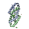
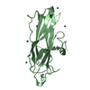
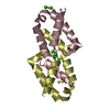
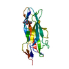
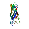
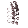
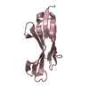
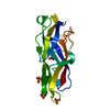
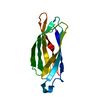
| Title | Crystal structure of catalytic domain of Anhydrobiosis-related Mn-dependent Peroxidase (AMNP) from Ramazzottius varieornatus (Mn2+-bound form) | ||||||
 Components Components | AMNP/g12777 | ||||||
 Keywords Keywords | OXIDOREDUCTASE / peroxidase / Manganese / tardigrade | ||||||
| Function / homology | : / Conserved secreted protein Function and homology information Function and homology information | ||||||
| Biological species |  | ||||||
| Method |  X-RAY DIFFRACTION / SYNCHROTRON / MOLECULAR REPLACEMENT / Resolution: 2.3 Å X-RAY DIFFRACTION / SYNCHROTRON / MOLECULAR REPLACEMENT / Resolution: 2.3 Å | ||||||
 Authors Authors | Yoshida, Y. / Satoh, T. / Ota, C. / Tanaka, S. / Horikawa, D.D. / Tomita, M. / Kato, K. / Arakawa, K. | ||||||
| Funding support |  Japan, 1items Japan, 1items
| ||||||
 Citation Citation | Journal: Bmc Genomics / Year: 2022 Title: Time-series transcriptomic screening of factors contributing to the cross-tolerance to UV radiation and anhydrobiosis in tardigrades. Authors: Yoshida, Y. / Satoh, T. / Ota, C. / Tanaka, S. / Horikawa, D.D. / Tomita, M. / Kato, K. / Arakawa, K. | ||||||
| History |
|
- Structure visualization
| Structure viewer | Molecule: MolmilJmol/JSmol |
|---|
- Downloads & links
-Download
| PDBx/mmCIF format | 7dbt.cif.gz | 76.8 KB | Display | PDBx/mmCIF format |
|---|---|---|---|---|
| PDB format | pdb7dbt.ent.gz | 54.8 KB | Display | PDB format |
| PDBx/mmJSON format | 7dbt.json.gz | Tree view | PDBx/mmJSON format | |
| Others |  Other downloads Other downloads |
-Validation report
| Summary document | 7dbt_validation.pdf.gz | 2 MB | Display | wwPDB validaton report |
|---|---|---|---|---|
| Full document | 7dbt_full_validation.pdf.gz | 2 MB | Display | |
| Data in XML | 7dbt_validation.xml.gz | 14.6 KB | Display | |
| Data in CIF | 7dbt_validation.cif.gz | 19.5 KB | Display | |
| Arichive directory | https://data.pdbj.org/pub/pdb/validation_reports/db/7dbtftp://data.pdbj.org/pub/pdb/validation_reports/db/7dbt | HTTPS FTP |
-Related structure data
| Related structure data |  7dbuSC S: Starting model for refinement C: citing same article ( |
|---|---|
| Similar structure data |
-Links
 PDBj
PDBj- Assembly
| Deposited unit | 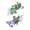
| ||||||||
|---|---|---|---|---|---|---|---|---|---|
| 1 | 
| ||||||||
| 2 | 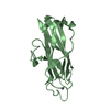
| ||||||||
| Unit cell |
|
-Components
| #1: Protein | Mass: 18743.023 Da / Num. of mol.: 2 Source method: isolated from a genetically manipulated source Source: (gene. exp.) Gene: RvY_12637-1, RvY_12637.1, RvY_12637 / Plasmid: pET28b / Production host:  #2: Chemical | ChemComp-MN /   Mass: 54.938 Da / Num. of mol.: 4 / Source method: obtained synthetically / Formula: Mn / Feature type: SUBJECT OF INVESTIGATION Mass: 54.938 Da / Num. of mol.: 4 / Source method: obtained synthetically / Formula: Mn / Feature type: SUBJECT OF INVESTIGATION#3: Water | ChemComp-HOH / |  Mass: 18.015 Da / Num. of mol.: 100 / Source method: isolated from a natural source / Formula: H2O Mass: 18.015 Da / Num. of mol.: 100 / Source method: isolated from a natural source / Formula: H2OHas ligand of interest | Y | |
|---|
-Experimental details
-Experiment
| Experiment | Method: X-RAY DIFFRACTION / Number of used crystals: 1 |
|---|
- Sample preparation
| Crystal | Density Matthews: 2.12 Å3/Da / Density % sol: 41.93 % |
|---|---|
| Crystal grow | Temperature: 293 K / Method: vapor diffusion, hanging drop / pH: 7.5 Details: Crystallization: 10% PEG3350, 100 mM imidazole-HCl buffer (pH 7.5), 300 mM zinc acetate, and 50 mM sodium fluoride Soaking: 11% PEG3350, 100 mM imidazole-HCl buffer (pH 7.5), 50 mM manganese ...Details: Crystallization: 10% PEG3350, 100 mM imidazole-HCl buffer (pH 7.5), 300 mM zinc acetate, and 50 mM sodium fluoride Soaking: 11% PEG3350, 100 mM imidazole-HCl buffer (pH 7.5), 50 mM manganese chloride, and 50 mM sodium fluoride |
-Data collection
| Diffraction | Mean temperature: 95 K / Serial crystal experiment: N |
|---|---|
| Diffraction source | Source: SYNCHROTRON / Site: SPring-8 / Beamline: BL44XU / Wavelength: 1.25 Å |
| Detector | Type: DECTRIS EIGER X 16M / Detector: PIXEL / Date: Jul 20, 2020 |
| Radiation | Monochromator: Si(111) / Protocol: SINGLE WAVELENGTH / Monochromatic (M) / Laue (L): M / Scattering type: x-ray |
| Radiation wavelength | Wavelength: 1.25 Å / Relative weight: 1 |
| Reflection | Resolution: 2.3→42.09 Å / Num. obs: 14127 / % possible obs: 98.6 % / Redundancy: 4.4 % / CC1/2: 0.987 / Rmerge(I) obs: 0.16 / Net I/σ(I): 4.6 |
| Reflection shell | Resolution: 2.3→2.38 Å / Redundancy: 4.2 % / Rmerge(I) obs: 0.547 / Mean I/σ(I) obs: 1.7 / Num. unique obs: 1337 / CC1/2: 0.866 / % possible all: 48.4 |
- Processing
| Software |
| ||||||||||||||||||||||||||||||||||||||||||
|---|---|---|---|---|---|---|---|---|---|---|---|---|---|---|---|---|---|---|---|---|---|---|---|---|---|---|---|---|---|---|---|---|---|---|---|---|---|---|---|---|---|---|---|
| Refinement | Method to determine structure: MOLECULAR REPLACEMENT Starting model: 7DBU Resolution: 2.3→19.93 Å / SU ML: 0.3 / Cross valid method: FREE R-VALUE / σ(F): 1.34 / Phase error: 42 / Stereochemistry target values: ML
| ||||||||||||||||||||||||||||||||||||||||||
| Solvent computation | Shrinkage radii: 0.9 Å / VDW probe radii: 1.11 Å / Solvent model: FLAT BULK SOLVENT MODEL | ||||||||||||||||||||||||||||||||||||||||||
| Refinement step | Cycle: LAST / Resolution: 2.3→19.93 Å
| ||||||||||||||||||||||||||||||||||||||||||
| Refine LS restraints |
| ||||||||||||||||||||||||||||||||||||||||||
| LS refinement shell |
|
