 Movie
Movie Controller

[English] 日本語
 Yorodumi
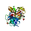
- PDB-7d8d: The crystal structure of ScNTM1 in complex with SAH and Rps25a he... -
+ Open data

- Basic information
| Entry | Database: PDB / ID: 7d8d | |||||||||
|---|---|---|---|---|---|---|---|---|---|---|
| Title | The crystal structure of ScNTM1 in complex with SAH and Rps25a hexapeptide | |||||||||
 Components Components |
| |||||||||
 Keywords Keywords | TRANSFERASE / ScNTM1 / N-terminal methylation / methyltransferase / Saccharomyces cerevisiae / substrate binding pocket | |||||||||
| Function / homology |  Function and homology information Function and homology informationN-terminal peptidyl-proline dimethylation / protein N-terminal methyltransferase / N-terminal protein N-methyltransferase activity / translational readthrough / Formation of the ternary complex, and subsequently, the 43S complex / Translation initiation complex formation / Ribosomal scanning and start codon recognition / SRP-dependent cotranslational protein targeting to membrane / GTP hydrolysis and joining of the 60S ribosomal subunit / Formation of a pool of free 40S subunits ...N-terminal peptidyl-proline dimethylation / protein N-terminal methyltransferase / N-terminal protein N-methyltransferase activity / translational readthrough / Formation of the ternary complex, and subsequently, the 43S complex / Translation initiation complex formation / Ribosomal scanning and start codon recognition / SRP-dependent cotranslational protein targeting to membrane / GTP hydrolysis and joining of the 60S ribosomal subunit / Formation of a pool of free 40S subunits / Nonsense Mediated Decay (NMD) independent of the Exon Junction Complex (EJC) / Nonsense Mediated Decay (NMD) enhanced by the Exon Junction Complex (EJC) / L13a-mediated translational silencing of Ceruloplasmin expression / cytosolic small ribosomal subunit / cytoplasmic translation / structural constituent of ribosome / cytosol / cytoplasm Similarity search - Function | |||||||||
| Biological species |  | |||||||||
| Method |  X-RAY DIFFRACTION / SYNCHROTRON / MOLECULAR REPLACEMENT / Resolution: 1.05 Å X-RAY DIFFRACTION / SYNCHROTRON / MOLECULAR REPLACEMENT / Resolution: 1.05 Å | |||||||||
 Authors Authors | Zhang, H.Y. / Yue, J. | |||||||||
| Funding support |  China, 2items China, 2items
| |||||||||
 Citation Citation | Journal: Crystallography Reports / Year: 2021 Title: Structural Basis for Peptide Binding of Alpha-N Terminal Methyltransferase from Saccharomyces cerevisiae Authors: Zhang, H.Y. / Kuang, Z. / Xue, L. / Yue, J. / Khan, M.H. / Zhu, Z. / Niu, L. | |||||||||
| History |
|
- Structure visualization
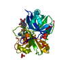
| Structure viewer | Molecule: MolmilJmol/JSmol |
|---|
- Downloads & links
-Download
| PDBx/mmCIF format | 7d8d.cif.gz | 199.7 KB | Display | PDBx/mmCIF format |
|---|---|---|---|---|
| PDB format | pdb7d8d.ent.gz | 131.1 KB | Display | PDB format |
| PDBx/mmJSON format | 7d8d.json.gz | Tree view | PDBx/mmJSON format | |
| Others |  Other downloads Other downloads |
-Validation report
| Arichive directory | https://data.pdbj.org/pub/pdb/validation_reports/d8/7d8dftp://data.pdbj.org/pub/pdb/validation_reports/d8/7d8d | HTTPS FTP |
|---|
-Related structure data
| Related structure data | 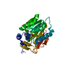 7d8fC  2ex4S S: Starting model for refinement C: citing same article ( |
|---|---|
| Similar structure data |


-Links
 PDBj
PDBj


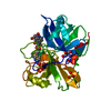
- Assembly
| Deposited unit | 
| ||||||||||||
|---|---|---|---|---|---|---|---|---|---|---|---|---|---|
| 1 |
| ||||||||||||
| Unit cell |
|
-Components
| #1: Protein | Mass: 26098.775 Da / Num. of mol.: 1 Source method: isolated from a genetically manipulated source Source: (gene. exp.) Gene: TAE1, NTM1, YBR261C, YBR1729 / Production host:  References: UniProt: P38340, protein N-terminal methyltransferase |
|---|---|
| #2: Protein/peptide | Mass: 710.840 Da / Num. of mol.: 1 / Source method: obtained synthetically / Source: (synth.) |
| #3: Chemical | ChemComp-SAH /   Type: L-peptide linking / Mass: 384.411 Da / Num. of mol.: 1 / Source method: isolated from a natural source / Formula: C14H20N6O5S / Feature type: SUBJECT OF INVESTIGATION Type: L-peptide linking / Mass: 384.411 Da / Num. of mol.: 1 / Source method: isolated from a natural source / Formula: C14H20N6O5S / Feature type: SUBJECT OF INVESTIGATION |
| #4: Water | ChemComp-HOH /  Mass: 18.015 Da / Num. of mol.: 327 / Source method: isolated from a natural source / Formula: H2O Mass: 18.015 Da / Num. of mol.: 327 / Source method: isolated from a natural source / Formula: H2O |
| Has ligand of interest | Y |
-Experimental details
-Experiment
| Experiment | Method: X-RAY DIFFRACTION / Number of used crystals: 1 |
|---|
- Sample preparation
| Crystal | Density Matthews: 2.32 Å3/Da / Density % sol: 47 % |
|---|---|
| Crystal grow | Temperature: 291 K / Method: vapor diffusion, sitting drop / pH: 6.5 Details: 0.2M magnesium chloride hexahydrate,0.1M Bis-Tris(pH 6.5) and 25% PEG3350 PH range: 6.0-7.0 |
-Data collection
| Diffraction | Mean temperature: 100 K / Serial crystal experiment: N |
|---|---|
| Diffraction source | Source: SYNCHROTRON / Site: SSRF / Beamline: BL17U / Wavelength: 0.97891 Å |
| Detector | Type: MARMOSAIC 300 mm CCD / Detector: CCD / Date: Jan 16, 2020 |
| Radiation | Protocol: SINGLE WAVELENGTH / Monochromatic (M) / Laue (L): M / Scattering type: x-ray |
| Radiation wavelength | Wavelength: 0.97891 Å / Relative weight: 1 |
| Reflection | Resolution: 0.999→50 Å / Num. obs: 130264 / % possible obs: 93 % / Redundancy: 8.6 % / Biso Wilson estimate: 6.32 Å2 / CC1/2: 0.998 / Rpim(I) all: 0.031 / Rrim(I) all: 0.094 / Net I/σ(I): 38.8 |
| Reflection shell | Resolution: 1→1.02 Å / Num. unique obs: 3404 / CC1/2: 0.032 / % possible all: 49 |
- Processing
| Software |
| |||||||||||||||||||||||||||||||||||||||||||||||||||||||||||||||||||||||||||||||||||||||||||||||||||||||||||||||||||||||||||||||||||||||||||||||||||
|---|---|---|---|---|---|---|---|---|---|---|---|---|---|---|---|---|---|---|---|---|---|---|---|---|---|---|---|---|---|---|---|---|---|---|---|---|---|---|---|---|---|---|---|---|---|---|---|---|---|---|---|---|---|---|---|---|---|---|---|---|---|---|---|---|---|---|---|---|---|---|---|---|---|---|---|---|---|---|---|---|---|---|---|---|---|---|---|---|---|---|---|---|---|---|---|---|---|---|---|---|---|---|---|---|---|---|---|---|---|---|---|---|---|---|---|---|---|---|---|---|---|---|---|---|---|---|---|---|---|---|---|---|---|---|---|---|---|---|---|---|---|---|---|---|---|---|---|---|
| Refinement | Method to determine structure: MOLECULAR REPLACEMENT Starting model: 2EX4 Resolution: 1.05→27.21 Å / SU ML: 0.0649 / Cross valid method: FREE R-VALUE / σ(F): 1.34 / Phase error: 10.4271 Stereochemistry target values: GeoStd + Monomer Library + CDL v1.2
| |||||||||||||||||||||||||||||||||||||||||||||||||||||||||||||||||||||||||||||||||||||||||||||||||||||||||||||||||||||||||||||||||||||||||||||||||||
| Solvent computation | Shrinkage radii: 0.9 Å / VDW probe radii: 1.11 Å / Solvent model: FLAT BULK SOLVENT MODEL | |||||||||||||||||||||||||||||||||||||||||||||||||||||||||||||||||||||||||||||||||||||||||||||||||||||||||||||||||||||||||||||||||||||||||||||||||||
| Displacement parameters | Biso mean: 10.37 Å2 | |||||||||||||||||||||||||||||||||||||||||||||||||||||||||||||||||||||||||||||||||||||||||||||||||||||||||||||||||||||||||||||||||||||||||||||||||||
| Refinement step | Cycle: LAST / Resolution: 1.05→27.21 Å
| |||||||||||||||||||||||||||||||||||||||||||||||||||||||||||||||||||||||||||||||||||||||||||||||||||||||||||||||||||||||||||||||||||||||||||||||||||
| Refine LS restraints |
| |||||||||||||||||||||||||||||||||||||||||||||||||||||||||||||||||||||||||||||||||||||||||||||||||||||||||||||||||||||||||||||||||||||||||||||||||||
| LS refinement shell |
|
