 Movie
Movie Controller


+ Open data

- Basic information
| Entry | Database: PDB / ID: 7d6l | ||||||
|---|---|---|---|---|---|---|---|
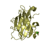
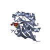
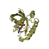
| Title | Crystal structure of Trx2 from D. radiodurans R1 | ||||||
 Components Components | Thioredoxin 2 | ||||||
 Keywords Keywords | OXIDOREDUCTASE / antioxidant / thioredoxin / redox control / cysteine thiol-disulfide exchange | ||||||
| Function / homology |  Function and homology information Function and homology informationprotein-disulfide reductase activity / cell redox homeostasis / metal ion binding / cytoplasm / cytosol Similarity search - Function | ||||||
| Biological species |  Deinococcus radiodurans R1 (radioresistant) Deinococcus radiodurans R1 (radioresistant) | ||||||
| Method |  X-RAY DIFFRACTION / SYNCHROTRON / MOLECULAR REPLACEMENT / molecular replacement / Resolution: 1.947 Å X-RAY DIFFRACTION / SYNCHROTRON / MOLECULAR REPLACEMENT / molecular replacement / Resolution: 1.947 Å | ||||||
 Authors Authors | Kim, M.-K. / Zhang, J. / Zhao, L. | ||||||
| Funding support |  Korea, Republic Of, 1items Korea, Republic Of, 1items
| ||||||
 Citation Citation | Journal: Antioxidants / Year: 2021 Title: Structural and Biochemical Characterization of Thioredoxin-2 from Deinococcus radiodurans. Authors: Kim, M.-K. / Zhao, L. / Jeong, S. / Zhang, J. / Jung, J.-H. / Seo, H.S. / Choi, J.- / Lim, S. | ||||||
| History |
|
- Structure visualization
| Structure viewer | Molecule: MolmilJmol/JSmol |
|---|
- Downloads & links
-Download
| PDBx/mmCIF format | 7d6l.cif.gz | 71.3 KB | Display | PDBx/mmCIF format |
|---|---|---|---|---|
| PDB format | pdb7d6l.ent.gz | 51 KB | Display | PDB format |
| PDBx/mmJSON format | 7d6l.json.gz | Tree view | PDBx/mmJSON format | |
| Others |  Other downloads Other downloads |
-Validation report
| Arichive directory | https://data.pdbj.org/pub/pdb/validation_reports/d6/7d6lftp://data.pdbj.org/pub/pdb/validation_reports/d6/7d6l | HTTPS FTP |
|---|
-Related structure data
| Related structure data |  1v98S S: Starting model for refinement |
|---|---|
| Similar structure data |


-Links
 PDBj
PDBj

- Assembly
| Deposited unit | 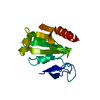
| ||||||||
|---|---|---|---|---|---|---|---|---|---|
| 1 |
| ||||||||
| Unit cell |
|
-Components
| #1: Protein | Mass: 16383.777 Da / Num. of mol.: 1 Source method: isolated from a genetically manipulated source Source: (gene. exp.) Deinococcus radiodurans R1 (radioresistant)Strain: R1 / Gene: DR_A0164 / Production host: |
|---|---|
| #2: Chemical | ChemComp-ZN /   Mass: 65.409 Da / Num. of mol.: 1 / Source method: obtained synthetically / Formula: Zn / Feature type: SUBJECT OF INVESTIGATION Mass: 65.409 Da / Num. of mol.: 1 / Source method: obtained synthetically / Formula: Zn / Feature type: SUBJECT OF INVESTIGATION |
| #3: Water | ChemComp-HOH /  Mass: 18.015 Da / Num. of mol.: 74 / Source method: isolated from a natural source / Formula: H2O Mass: 18.015 Da / Num. of mol.: 74 / Source method: isolated from a natural source / Formula: H2O |
| Has ligand of interest | Y |
| Has protein modification | Y |
-Experimental details
-Experiment
| Experiment | Method: X-RAY DIFFRACTION / Number of used crystals: 1 |
|---|
- Sample preparation
| Crystal | Density Matthews: 3.24 Å3/Da / Density % sol: 62.02 % |
|---|---|
| Crystal grow | Temperature: 295 K / Method: microbatch / Details: PEG 3350, Bis-Tris |
-Data collection
| Diffraction | Mean temperature: 100 K / Serial crystal experiment: N |
|---|---|
| Diffraction source | Source: SYNCHROTRON / Site: PAL/PLS / Beamline: 5C (4A) / Wavelength: 0.9794 Å |
| Detector | Type: DECTRIS EIGER X 9M / Detector: PIXEL / Date: Oct 3, 2018 |
| Radiation | Protocol: SINGLE WAVELENGTH / Monochromatic (M) / Laue (L): M / Scattering type: x-ray |
| Radiation wavelength | Wavelength: 0.9794 Å / Relative weight: 1 |
| Reflection | Resolution: 1.947→50 Å / Num. obs: 14566 / % possible obs: 94.9 % / Redundancy: 5.1 % / CC1/2: 0.993 / Rpim(I) all: 0.03 / Rrim(I) all: 0.072 / Net I/σ(I): 50.779 |
| Reflection shell | Resolution: 1.947→1.99 Å / Mean I/σ(I) obs: 11.628 / Num. unique obs: 685 / CC1/2: 0.971 / Rpim(I) all: 0.118 / Rrim(I) all: 0.262 |
-Phasing
| Phasing | Method: molecular replacement |
|---|
- Processing
| Software |
| ||||||||||||||||||||||||||||||||||||||||||||||||||||||||||||||||||
|---|---|---|---|---|---|---|---|---|---|---|---|---|---|---|---|---|---|---|---|---|---|---|---|---|---|---|---|---|---|---|---|---|---|---|---|---|---|---|---|---|---|---|---|---|---|---|---|---|---|---|---|---|---|---|---|---|---|---|---|---|---|---|---|---|---|---|---|
| Refinement | Method to determine structure: MOLECULAR REPLACEMENT Starting model: 1v98 Resolution: 1.947→18.935 Å / SU ML: 0.23 / Cross valid method: THROUGHOUT / σ(F): 1.54 / Phase error: 22.99 / Stereochemistry target values: ML
| ||||||||||||||||||||||||||||||||||||||||||||||||||||||||||||||||||
| Solvent computation | Shrinkage radii: 0.9 Å / VDW probe radii: 1.11 Å / Solvent model: FLAT BULK SOLVENT MODEL | ||||||||||||||||||||||||||||||||||||||||||||||||||||||||||||||||||
| Displacement parameters | Biso max: 108.28 Å2 / Biso mean: 38.3938 Å2 / Biso min: 13.57 Å2 | ||||||||||||||||||||||||||||||||||||||||||||||||||||||||||||||||||
| Refinement step | Cycle: final / Resolution: 1.947→18.935 Å
| ||||||||||||||||||||||||||||||||||||||||||||||||||||||||||||||||||
| LS refinement shell | Refine-ID: X-RAY DIFFRACTION / Rfactor Rfree error: 0
| ||||||||||||||||||||||||||||||||||||||||||||||||||||||||||||||||||
| Refinement TLS params. | Method: refined / Origin x: 35.221 Å / Origin y: 50.7825 Å / Origin z: 6.1787 Å
| ||||||||||||||||||||||||||||||||||||||||||||||||||||||||||||||||||
| Refinement TLS group |
|
