 Movie
Movie Controller

[English] 日本語
 Yorodumi
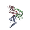
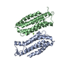
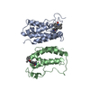
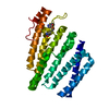
- PDB-7d2w: Crystal structure of PHIT/PHISTa-like subfamily PF3D7_1372300 pro... -
+ Open data

- Basic information
| Entry | Database: PDB / ID: 7d2w | ||||||
|---|---|---|---|---|---|---|---|
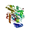
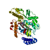
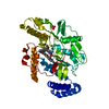
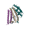
| Title | Crystal structure of PHIT/PHISTa-like subfamily PF3D7_1372300 protein from plasmodium falciparum | ||||||
 Components Components | PRESAN domain-containing protein | ||||||
 Keywords Keywords | PROTEIN BINDING / four-helix bundle structure | ||||||
| Function / homology | Exported protein, PHISTa/b/c, conserved domain, Plasmodium / Plasmodium RESA, N-terminal / Plasmodium RESA, N-terminal helical domain superfamily / Plasmodium RESA N-terminal / : / Plasmodium RESA N-terminal domain-containing protein Function and homology information Function and homology information | ||||||
| Biological species |  | ||||||
| Method |  X-RAY DIFFRACTION / SYNCHROTRON / SAD / Resolution: 1.97 Å X-RAY DIFFRACTION / SYNCHROTRON / SAD / Resolution: 1.97 Å | ||||||
 Authors Authors | Gao, Y. / Yang, B.L. | ||||||
| Funding support |  China, 1items China, 1items
| ||||||
 Citation Citation | Journal: To Be Published Title: Structural analysis of binding between Plasmodium falciparum PHIST and the parasite virulence factor PfEMP1 protein Authors: Yang, B.L. / Gao, Y. | ||||||
| History |
|
- Structure visualization
| Structure viewer | Molecule: MolmilJmol/JSmol |
|---|
- Downloads & links
-Download
| PDBx/mmCIF format | 7d2w.cif.gz | 156.3 KB | Display | PDBx/mmCIF format |
|---|---|---|---|---|
| PDB format | pdb7d2w.ent.gz | 102.3 KB | Display | PDB format |
| PDBx/mmJSON format | 7d2w.json.gz | Tree view | PDBx/mmJSON format | |
| Others |  Other downloads Other downloads |
-Validation report
| Summary document | 7d2w_validation.pdf.gz | 437.2 KB | Display | wwPDB validaton report |
|---|---|---|---|---|
| Full document | 7d2w_full_validation.pdf.gz | 438.2 KB | Display | |
| Data in XML | 7d2w_validation.xml.gz | 13.8 KB | Display | |
| Data in CIF | 7d2w_validation.cif.gz | 19.7 KB | Display | |
| Arichive directory | https://data.pdbj.org/pub/pdb/validation_reports/d2/7d2wftp://data.pdbj.org/pub/pdb/validation_reports/d2/7d2w | HTTPS FTP |
-Related structure data
| Similar structure data |
|---|
-Links
 PDBj
PDBj


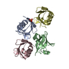
- Assembly
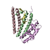
| Deposited unit | 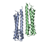
| ||||||||||||
|---|---|---|---|---|---|---|---|---|---|---|---|---|---|
| 1 |
| ||||||||||||
| Unit cell |
|
-Components
| #1: Protein | Mass: 22455.551 Da / Num. of mol.: 2 Source method: isolated from a genetically manipulated source Source: (gene. exp.) Gene: PF3D7_1372300 Production host:  References: UniProt: A0A143ZZR8 #2: Chemical | ChemComp-HG / |   Mass: 200.590 Da / Num. of mol.: 1 / Source method: obtained synthetically / Formula: Hg Mass: 200.590 Da / Num. of mol.: 1 / Source method: obtained synthetically / Formula: Hg#3: Water | ChemComp-HOH / |  Mass: 18.015 Da / Num. of mol.: 240 / Source method: isolated from a natural source / Formula: H2O Mass: 18.015 Da / Num. of mol.: 240 / Source method: isolated from a natural source / Formula: H2OHas ligand of interest | N | |
|---|
-Experimental details
-Experiment
| Experiment | Method: X-RAY DIFFRACTION / Number of used crystals: 1 |
|---|
- Sample preparation
| Crystal | Density Matthews: 1.99 Å3/Da / Density % sol: 38.27 % |
|---|---|
| Crystal grow | Temperature: 291 K / Method: vapor diffusion / Details: 16% PEG 8000, 50mM KH2PO4 |
-Data collection
| Diffraction | Mean temperature: 100 K / Serial crystal experiment: N |
|---|---|
| Diffraction source | Source: SYNCHROTRON / Site: SSRF / Beamline: BL19U1 / Wavelength: 1.0082 Å |
| Detector | Type: DECTRIS PILATUS3 S 6M / Detector: PIXEL / Date: May 31, 2018 |
| Radiation | Monochromator: M / Protocol: SINGLE WAVELENGTH / Monochromatic (M) / Laue (L): M / Scattering type: x-ray |
| Radiation wavelength | Wavelength: 1.0082 Å / Relative weight: 1 |
| Reflection | Resolution: 1.97→50 Å / Num. obs: 24930 / % possible obs: 100 % / Redundancy: 20 % / Biso Wilson estimate: 32.35 Å2 / CC1/2: 1 / Net I/σ(I): 45.45 |
| Reflection shell | Resolution: 1.97→2.04 Å / Redundancy: 20.5 % / Num. unique obs: 2472 / CC1/2: 0.972 / % possible all: 100 |
- Processing
| Software |
| |||||||||||||||||||||||||||||||||||||||||||||||||||||||||||||||||||||||||||||||||||||||||||||||||||||||||
|---|---|---|---|---|---|---|---|---|---|---|---|---|---|---|---|---|---|---|---|---|---|---|---|---|---|---|---|---|---|---|---|---|---|---|---|---|---|---|---|---|---|---|---|---|---|---|---|---|---|---|---|---|---|---|---|---|---|---|---|---|---|---|---|---|---|---|---|---|---|---|---|---|---|---|---|---|---|---|---|---|---|---|---|---|---|---|---|---|---|---|---|---|---|---|---|---|---|---|---|---|---|---|---|---|---|---|
| Refinement | Method to determine structure: SAD / Resolution: 1.97→40.43 Å / SU ML: 0.2055 / Cross valid method: FREE R-VALUE / σ(F): 0 / Phase error: 22.3592 / Stereochemistry target values: CDL v1.2
| |||||||||||||||||||||||||||||||||||||||||||||||||||||||||||||||||||||||||||||||||||||||||||||||||||||||||
| Solvent computation | Shrinkage radii: 0.9 Å / VDW probe radii: 1.11 Å / Solvent model: FLAT BULK SOLVENT MODEL | |||||||||||||||||||||||||||||||||||||||||||||||||||||||||||||||||||||||||||||||||||||||||||||||||||||||||
| Displacement parameters | Biso mean: 41.48 Å2 | |||||||||||||||||||||||||||||||||||||||||||||||||||||||||||||||||||||||||||||||||||||||||||||||||||||||||
| Refinement step | Cycle: LAST / Resolution: 1.97→40.43 Å
| |||||||||||||||||||||||||||||||||||||||||||||||||||||||||||||||||||||||||||||||||||||||||||||||||||||||||
| Refine LS restraints |
| |||||||||||||||||||||||||||||||||||||||||||||||||||||||||||||||||||||||||||||||||||||||||||||||||||||||||
| LS refinement shell |
| |||||||||||||||||||||||||||||||||||||||||||||||||||||||||||||||||||||||||||||||||||||||||||||||||||||||||
| Refinement TLS params. | Method: refined / Origin x: 8.04585106279 Å / Origin y: 38.8152267928 Å / Origin z: 38.4751052902 Å
| |||||||||||||||||||||||||||||||||||||||||||||||||||||||||||||||||||||||||||||||||||||||||||||||||||||||||
| Refinement TLS group | Selection details: all |
