[English] 日本語
 Yorodumi
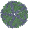
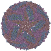
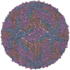
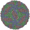
- PDB-7d0l: The major capsid of Omono River virus (strain:LZ), protrusion-fre... -
+ Open data
- Basic information
| Entry | Database: PDB / ID: 7d0l | ||||||
|---|---|---|---|---|---|---|---|
| Title | The major capsid of Omono River virus (strain:LZ), protrusion-free status. | ||||||
 Components Components | Capsid protein | ||||||
 Keywords Keywords | VIRUS / Totiviridae / major capsid / viral protein | ||||||
| Function / homology | Double-stranded RNA binding motif / Double-stranded RNA binding motif / Double stranded RNA-binding domain (dsRBD) profile. / Double-stranded RNA-binding domain / RNA binding / Main capsid protein Function and homology information Function and homology information | ||||||
| Biological species |  Omono River virus Omono River virus | ||||||
| Method | ELECTRON MICROSCOPY / single particle reconstruction / cryo EM / Resolution: 2.95 Å | ||||||
 Authors Authors | Shao, Q. / Jia, X. / Gao, Y. / Liu, Z. | ||||||
| Funding support |  China, 1items China, 1items
| ||||||
 Citation Citation |  Journal: PLoS Pathog / Year: 2021 Journal: PLoS Pathog / Year: 2021Title: Cryo-EM reveals a previously unrecognized structural protein of a dsRNA virus implicated in its extracellular transmission. Authors: Qianqian Shao / Xudong Jia / Yuanzhu Gao / Zhe Liu / Huan Zhang / Qiqi Tan / Xin Zhang / Huiqiong Zhou / Yinyin Li / De Wu / Qinfen Zhang /  Abstract: Mosquito viruses cause unpredictable outbreaks of disease. Recently, several unassigned viruses isolated from mosquitoes, including the Omono River virus (OmRV), were identified as totivirus-like ...Mosquito viruses cause unpredictable outbreaks of disease. Recently, several unassigned viruses isolated from mosquitoes, including the Omono River virus (OmRV), were identified as totivirus-like viruses, with features similar to those of the Totiviridae family. Most reported members of this family infect fungi or protozoans and lack an extracellular life cycle stage. Here, we identified a new strain of OmRV and determined high-resolution structures for this virus using single-particle cryo-electron microscopy. The structures feature an unexpected protrusion at the five-fold vertex of the capsid. Disassociation of the protrusion could result in several conformational changes in the major capsid. All these structures, together with some biological results, suggest the protrusions' associations with the extracellular transmission of OmRV. | ||||||
| History |
|
- Structure visualization
| Movie |
 Movie viewer Movie viewer |
|---|---|
| Structure viewer | Molecule:  Molmil Molmil Jmol/JSmol Jmol/JSmol |
- Downloads & links
- Download
| PDBx/mmCIF format |  7d0l.cif.gz 7d0l.cif.gz | 305.6 KB | Display |  PDBx/mmCIF format PDBx/mmCIF format |
|---|---|---|---|---|
| PDB format |  pdb7d0l.ent.gz pdb7d0l.ent.gz | 234.5 KB | Display |  PDB format PDB format |
| PDBx/mmJSON format |  7d0l.json.gz 7d0l.json.gz | Tree view |  PDBx/mmJSON format PDBx/mmJSON format | |
| Others |  Other downloads Other downloads |
-Validation report
| Arichive directory |  https://data.pdbj.org/pub/pdb/validation_reports/d0/7d0l https://data.pdbj.org/pub/pdb/validation_reports/d0/7d0l ftp://data.pdbj.org/pub/pdb/validation_reports/d0/7d0l ftp://data.pdbj.org/pub/pdb/validation_reports/d0/7d0l | HTTPS FTP |
|---|
-Related structure data
| Related structure data |  30538MC  7cz6C  7d0kC M: map data used to model this data C: citing same article ( |
|---|---|
| Similar structure data |
- Links
Links
- Assembly
| Deposited unit | 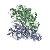
|
|---|---|
| 1 | x 60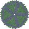
|
| 2 |
|
| 3 | x 5
|
| 4 | x 6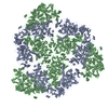
|
| 5 | 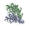
|
| Symmetry | Point symmetry: (Schoenflies symbol: I (icosahedral)) |
- Components
| #1: Protein | Mass: 96860.992 Da / Num. of mol.: 2 / Source method: isolated from a natural source / Source: (natural)  Omono River virus / Cell line: C6/36 / Strain: LZ / References: UniProt: A0A7M3VBX7*PLUS Omono River virus / Cell line: C6/36 / Strain: LZ / References: UniProt: A0A7M3VBX7*PLUS |
|---|
-Experimental details
-Experiment
| Experiment | Method: ELECTRON MICROSCOPY |
|---|---|
| EM experiment | Aggregation state: PARTICLE / 3D reconstruction method: single particle reconstruction |
- Sample preparation
| Component | Name: Major capsid dimer of OmRV-LZ (protrusion-free status) Type: VIRUS / Entity ID: all / Source: NATURAL | |||||||||||||||||||||||||
|---|---|---|---|---|---|---|---|---|---|---|---|---|---|---|---|---|---|---|---|---|---|---|---|---|---|---|
| Molecular weight | Experimental value: NO | |||||||||||||||||||||||||
| Source (natural) | Organism:  Omono River virus / Strain: LZ Omono River virus / Strain: LZ | |||||||||||||||||||||||||
| Details of virus | Empty: NO / Enveloped: NO / Isolate: STRAIN / Type: VIRION | |||||||||||||||||||||||||
| Natural host | Organism: Culex quinquefasciatus | |||||||||||||||||||||||||
| Virus shell | Name: Major capsid / Diameter: 450 nm / Triangulation number (T number): 1 | |||||||||||||||||||||||||
| Buffer solution | pH: 7.4 | |||||||||||||||||||||||||
| Buffer component |
| |||||||||||||||||||||||||
| Specimen | Embedding applied: NO / Shadowing applied: NO / Staining applied: NO / Vitrification applied: YES | |||||||||||||||||||||||||
| Specimen support | Grid material: COPPER / Grid mesh size: 300 divisions/in. / Grid type: Quantifoil R1.2/1.3 | |||||||||||||||||||||||||
| Vitrification | Instrument: FEI VITROBOT MARK IV / Cryogen name: ETHANE / Humidity: 100 % |
- Electron microscopy imaging
| Experimental equipment |  Model: Titan Krios / Image courtesy: FEI Company |
|---|---|
| Microscopy | Model: FEI TITAN KRIOS |
| Electron gun | Electron source:  FIELD EMISSION GUN / Accelerating voltage: 300 kV / Illumination mode: FLOOD BEAM FIELD EMISSION GUN / Accelerating voltage: 300 kV / Illumination mode: FLOOD BEAM |
| Electron lens | Mode: BRIGHT FIELD / Nominal magnification: 75000 X / Nominal defocus max: 2500 nm / Nominal defocus min: 1000 nm / Alignment procedure: NONE |
| Specimen holder | Cryogen: NITROGEN |
| Image recording | Electron dose: 39 e/Å2 / Detector mode: COUNTING / Film or detector model: FEI FALCON III (4k x 4k) |
- Processing
| EM software |
| ||||||||||||||||||||||||||||||||||||
|---|---|---|---|---|---|---|---|---|---|---|---|---|---|---|---|---|---|---|---|---|---|---|---|---|---|---|---|---|---|---|---|---|---|---|---|---|---|
| CTF correction | Type: PHASE FLIPPING AND AMPLITUDE CORRECTION | ||||||||||||||||||||||||||||||||||||
| Particle selection | Num. of particles selected: 96668 Details: Subtracted from every five-fold vertexes from 96668 virus particles. Each virus contains 12 five-fold vertexes. 96668x12=1160016. | ||||||||||||||||||||||||||||||||||||
| 3D reconstruction | Resolution: 2.95 Å / Resolution method: FSC 0.143 CUT-OFF / Num. of particles: 15240 / Symmetry type: POINT | ||||||||||||||||||||||||||||||||||||
| Atomic model building | Protocol: AB INITIO MODEL / Space: REAL |
 Movie
Movie Controller





 PDBj
PDBj
