 Movie
Movie Controller


+ Open data

- Basic information
| Entry | Database: PDB / ID: 7cuj | ||||||
|---|---|---|---|---|---|---|---|
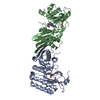
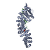
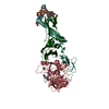
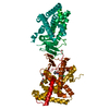
| Title | Crystal structure of fission yeast Ccq1 and Tpz1 | ||||||
 Components Components |
| ||||||
 Keywords Keywords | DNA BINDING PROTEIN / telomere | ||||||
| Function / homology |  Function and homology information Function and homology informationtelomere-telomerase complex assembly / subtelomeric heterochromatin / telomere cap complex / chromosome, telomeric repeat region / meiotic chromosome segregation / meiotic telomere clustering / shelterin complex / telomere capping / telomerase holoenzyme complex / protein localization to chromosome, telomeric region ...telomere-telomerase complex assembly / subtelomeric heterochromatin / telomere cap complex / chromosome, telomeric repeat region / meiotic chromosome segregation / meiotic telomere clustering / shelterin complex / telomere capping / telomerase holoenzyme complex / protein localization to chromosome, telomeric region / telomeric DNA binding / telomere maintenance via telomerase / telomere maintenance / molecular adaptor activity / nucleoplasm / nucleus Similarity search - Function | ||||||
| Biological species |  | ||||||
| Method |  X-RAY DIFFRACTION / SYNCHROTRON / SAD / Resolution: 2.4 Å X-RAY DIFFRACTION / SYNCHROTRON / SAD / Resolution: 2.4 Å | ||||||
 Authors Authors | Sun, H. / Wu, Z. / Wu, J. / Lei, M. | ||||||
 Citation Citation | Journal: Plos Genet. / Year: 2022 Title: Structural insights into Pot1-ssDNA, Pot1-Tpz1 and Tpz1-Ccq1 Interactions within fission yeast shelterin complex. Authors: Sun, H. / Wu, Z. / Zhou, Y. / Lu, Y. / Lu, H. / Chen, H. / Shi, S. / Zeng, Z. / Wu, J. / Lei, M. | ||||||
| History |
|
- Structure visualization
| Structure viewer | Molecule: MolmilJmol/JSmol |
|---|
- Downloads & links
-Download
| PDBx/mmCIF format | 7cuj.cif.gz | 254.7 KB | Display | PDBx/mmCIF format |
|---|---|---|---|---|
| PDB format | pdb7cuj.ent.gz | 207.1 KB | Display | PDB format |
| PDBx/mmJSON format | 7cuj.json.gz | Tree view | PDBx/mmJSON format | |
| Others |  Other downloads Other downloads |
-Validation report
| Summary document | 7cuj_validation.pdf.gz | 460.3 KB | Display | wwPDB validaton report |
|---|---|---|---|---|
| Full document | 7cuj_full_validation.pdf.gz | 469 KB | Display | |
| Data in XML | 7cuj_validation.xml.gz | 22.4 KB | Display | |
| Data in CIF | 7cuj_validation.cif.gz | 30.5 KB | Display | |
| Arichive directory | https://data.pdbj.org/pub/pdb/validation_reports/cu/7cujftp://data.pdbj.org/pub/pdb/validation_reports/cu/7cuj | HTTPS FTP |
-Related structure data


-Links
 PDBj
PDBj
- Assembly
| Deposited unit | 
| ||||||||
|---|---|---|---|---|---|---|---|---|---|
| 1 |
| ||||||||
| Unit cell |
|
-Components
| #1: Protein | Mass: 35256.641 Da / Num. of mol.: 2 Source method: isolated from a genetically manipulated source Source: (gene. exp.) Strain: 972 / ATCC 24843 / Gene: ccq1, SPCC188.07 / Plasmid: pET28a / Production host:  #2: Protein/peptide | Mass: 5579.404 Da / Num. of mol.: 2 Source method: isolated from a genetically manipulated source Source: (gene. exp.) Strain: 972 / ATCC 24843 / Gene: tpz1, mug169, SPAC6F6.16c, SPAC6F6.18c / Plasmid: pGEX6P1 / Production host: #3: Water | ChemComp-HOH / |  Mass: 18.015 Da / Num. of mol.: 80 / Source method: isolated from a natural source / Formula: H2O Mass: 18.015 Da / Num. of mol.: 80 / Source method: isolated from a natural source / Formula: H2O |
|---|
-Experimental details
-Experiment
| Experiment | Method: X-RAY DIFFRACTION / Number of used crystals: 1 |
|---|
- Sample preparation
| Crystal | Density Matthews: 2.37 Å3/Da / Density % sol: 48.02 % / Mosaicity: 0.41 ° |
|---|---|
| Crystal grow | Temperature: 277 K / Method: evaporation / pH: 7.8 Details: 15% PEG4000, 200mM potassium chloride, 50mM magnesium chloride and 50mM Tris-HCl |
-Data collection
| Diffraction |
| |||||||||||||||||||||||||||||||||||||||||||||||||||||||||||||||||||||||||||||||||||||||||||||||||||
|---|---|---|---|---|---|---|---|---|---|---|---|---|---|---|---|---|---|---|---|---|---|---|---|---|---|---|---|---|---|---|---|---|---|---|---|---|---|---|---|---|---|---|---|---|---|---|---|---|---|---|---|---|---|---|---|---|---|---|---|---|---|---|---|---|---|---|---|---|---|---|---|---|---|---|---|---|---|---|---|---|---|---|---|---|---|---|---|---|---|---|---|---|---|---|---|---|---|---|---|---|
| Diffraction source |
| |||||||||||||||||||||||||||||||||||||||||||||||||||||||||||||||||||||||||||||||||||||||||||||||||||
| Detector |
| |||||||||||||||||||||||||||||||||||||||||||||||||||||||||||||||||||||||||||||||||||||||||||||||||||
| Radiation |
| |||||||||||||||||||||||||||||||||||||||||||||||||||||||||||||||||||||||||||||||||||||||||||||||||||
| Radiation wavelength |
| |||||||||||||||||||||||||||||||||||||||||||||||||||||||||||||||||||||||||||||||||||||||||||||||||||
| Reflection | Resolution: 2.4→50 Å / Num. obs: 29579 / % possible obs: 98.9 % / Redundancy: 4.4 % / Rmerge(I) obs: 0.058 / Rpim(I) all: 0.03 / Rrim(I) all: 0.066 / Χ2: 0.913 / Net I/σ(I): 8.1 / Num. measured all: 131622 | |||||||||||||||||||||||||||||||||||||||||||||||||||||||||||||||||||||||||||||||||||||||||||||||||||
| Reflection shell | Diffraction-ID: 1
|

- Processing
| Software |
| |||||||||||||||||||||||||||||||||||||||||||||||||||||||||||||||||||||||||||||||||||||||||||||||||||||||||||||||||||||||||||||
|---|---|---|---|---|---|---|---|---|---|---|---|---|---|---|---|---|---|---|---|---|---|---|---|---|---|---|---|---|---|---|---|---|---|---|---|---|---|---|---|---|---|---|---|---|---|---|---|---|---|---|---|---|---|---|---|---|---|---|---|---|---|---|---|---|---|---|---|---|---|---|---|---|---|---|---|---|---|---|---|---|---|---|---|---|---|---|---|---|---|---|---|---|---|---|---|---|---|---|---|---|---|---|---|---|---|---|---|---|---|---|---|---|---|---|---|---|---|---|---|---|---|---|---|---|---|---|
| Refinement | Method to determine structure: SAD / Resolution: 2.4→38.87 Å / Cor.coef. Fo:Fc: 0.938 / Cor.coef. Fo:Fc free: 0.892 / SU B: 19.516 / SU ML: 0.202 / Cross valid method: THROUGHOUT / σ(F): 0 / ESU R Free: 0.297 / Stereochemistry target values: MAXIMUM LIKELIHOOD / Details: U VALUES : WITH TLS ADDED
| |||||||||||||||||||||||||||||||||||||||||||||||||||||||||||||||||||||||||||||||||||||||||||||||||||||||||||||||||||||||||||||
| Solvent computation | Ion probe radii: 0.8 Å / Shrinkage radii: 0.8 Å / VDW probe radii: 1.2 Å / Solvent model: MASK | |||||||||||||||||||||||||||||||||||||||||||||||||||||||||||||||||||||||||||||||||||||||||||||||||||||||||||||||||||||||||||||
| Displacement parameters | Biso max: 158.5 Å2 / Biso mean: 54.986 Å2 / Biso min: 12.22 Å2
| |||||||||||||||||||||||||||||||||||||||||||||||||||||||||||||||||||||||||||||||||||||||||||||||||||||||||||||||||||||||||||||
| Refinement step | Cycle: final / Resolution: 2.4→38.87 Å
| |||||||||||||||||||||||||||||||||||||||||||||||||||||||||||||||||||||||||||||||||||||||||||||||||||||||||||||||||||||||||||||
| Refine LS restraints |
| |||||||||||||||||||||||||||||||||||||||||||||||||||||||||||||||||||||||||||||||||||||||||||||||||||||||||||||||||||||||||||||
| LS refinement shell | Resolution: 2.4→2.462 Å / Rfactor Rfree error: 0 / Total num. of bins used: 20
| |||||||||||||||||||||||||||||||||||||||||||||||||||||||||||||||||||||||||||||||||||||||||||||||||||||||||||||||||||||||||||||
| Refinement TLS params. | Method: refined / Refine-ID: X-RAY DIFFRACTION
| |||||||||||||||||||||||||||||||||||||||||||||||||||||||||||||||||||||||||||||||||||||||||||||||||||||||||||||||||||||||||||||
| Refinement TLS group |
|
