- PDB-7ctp: Crystal Structure of Human FAM129B/MINERVA/NIBAN2 -
+
Open data
ID or keywords:
Loading...
-
Basic information
Entry
Database: PDB / ID: 7ctp
Title
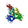
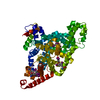
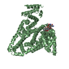
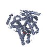
Crystal Structure of Human FAM129B/MINERVA/NIBAN2
Components
Protein Niban 2
Keywords
PROTEIN BINDING / PH domain / cytosol / plasma membrane
Function / homology
Function and homology information
gonadotropin secretion / negative regulation of DNA biosynthetic process / positive regulation of skeletal muscle fiber development / positive regulation of transcription regulatory region DNA binding / positive regulation of embryonic development / positive regulation of gene expression via chromosomal CpG island demethylation / negative regulation of vascular endothelial growth factor receptor signaling pathway / negative regulation of Notch signaling pathway / axon guidance / negative regulation of angiogenesis ...gonadotropin secretion / negative regulation of DNA biosynthetic process / positive regulation of skeletal muscle fiber development / positive regulation of transcription regulatory region DNA binding / positive regulation of embryonic development / positive regulation of gene expression via chromosomal CpG island demethylation / negative regulation of vascular endothelial growth factor receptor signaling pathway / negative regulation of Notch signaling pathway / axon guidance / negative regulation of angiogenesis / adherens junction / transcription coactivator activity / cell differentiation / cadherin binding / negative regulation of cell population proliferation / negative regulation of DNA-templated transcription / negative regulation of apoptotic process / positive regulation of DNA-templated transcription / extracellular exosome / nucleoplasm / nucleus / plasma membrane / cytoplasm / cytosol Similarity search - Function
In the structure databanks used in Yorodumi, some data are registered as the other names, "COVID-19 virus" and "2019-nCoV". Here are the details of the virus and the list of structure data.
Jan 31, 2019. EMDB accession codes are about to change! (news from PDBe EMDB page)
EMDB accession codes are about to change! (news from PDBe EMDB page)
The allocation of 4 digits for EMDB accession codes will soon come to an end. Whilst these codes will remain in use, new EMDB accession codes will include an additional digit and will expand incrementally as the available range of codes is exhausted. The current 4-digit format prefixed with “EMD-” (i.e. EMD-XXXX) will advance to a 5-digit format (i.e. EMD-XXXXX), and so on. It is currently estimated that the 4-digit codes will be depleted around Spring 2019, at which point the 5-digit format will come into force.
The EM Navigator/Yorodumi systems omit the EMD- prefix.
Related info.:Q: What is EMD? / ID/Accession-code notation in Yorodumi/EM Navigator
Yorodumi is a browser for structure data from EMDB, PDB, SASBDB, etc.
This page is also the successor to EM Navigator detail page, and also detail information page/front-end page for Omokage search.
The word "yorodu" (or yorozu) is an old Japanese word meaning "ten thousand". "mi" (miru) is to see.
Related info.:EMDB / PDB / SASBDB / Comparison of 3 databanks / Yorodumi Search / Aug 31, 2016. New EM Navigator & Yorodumi / Yorodumi Papers / Jmol/JSmol / Function and homology information / Changes in new EM Navigator and Yorodumi
 Movie
Movie Controller


 Open data
 Basic information
 Components
 Keywords
 Function and homology information
 Homo sapiens (human)
 X-RAY DIFFRACTION /
  Authors
 Korea, Republic Of, 4items
  Citation
 Structure visualization
 Downloads & links
 Other downloads


 PDBj
PDBj Assembly


 Mass: 92.094 Da / Num. of mol.: 3 / Source method: obtained synthetically / Formula: C3H8O3
 Mass: 18.015 Da / Num. of mol.: 269 / Source method: isolated from a natural source / Formula: H2O
 Sample preparation
 / Beamline: BL44XU / Wavelength: 1 Å
 Processing