 Movie
Movie Controller

[English] 日本語
 Yorodumi
- PDB-7ct4: Crystal structure of D-amino acid oxidase from Rasamsonia emerson... -
+ Open data

- Basic information
| Entry | Database: PDB / ID: 7ct4 | ||||||
|---|---|---|---|---|---|---|---|
| Title | Crystal structure of D-amino acid oxidase from Rasamsonia emersonii strain YA | ||||||
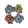
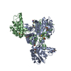
 Components Components | D-amino acid oxidase | ||||||
 Keywords Keywords | FLAVOPROTEIN / Oxidase / D-amino acid | ||||||
| Function / homology |  Function and homology information Function and homology informationD-amino acid metabolic process / D-amino-acid oxidase / D-amino-acid oxidase activity / D-amino acid catabolic process / nitrogen utilization / peroxisomal matrix / FAD binding Similarity search - Function | ||||||
| Biological species |  Talaromyces emersonii (fungus) Talaromyces emersonii (fungus) | ||||||
| Method |  X-RAY DIFFRACTION / SYNCHROTRON / MOLECULAR REPLACEMENT / Resolution: 2 Å X-RAY DIFFRACTION / SYNCHROTRON / MOLECULAR REPLACEMENT / Resolution: 2 Å | ||||||
 Authors Authors | Shimekake, Y. / Hirato, Y. / Okazaki, S. / Funabashi, R. / Goto, M. / Furuichi, T. / Suzuki, H. / Takahashi, S. | ||||||
 Citation Citation | Journal: Acta Crystallogr.,Sect.F / Year: 2020 Title: X-ray structure analysis of a unique D-amino-acid oxidase from the thermophilic fungus Rasamsonia emersonii strain YA. Authors: Shimekake, Y. / Hirato, Y. / Funabashi, R. / Okazaki, S. / Goto, M. / Furuichi, T. / Suzuki, H. / Kera, Y. / Takahashi, S. | ||||||
| History |
|
- Structure visualization
| Structure viewer | Molecule: MolmilJmol/JSmol |
|---|
- Downloads & links
-Download
| PDBx/mmCIF format | 7ct4.cif.gz | 279 KB | Display | PDBx/mmCIF format |
|---|---|---|---|---|
| PDB format | pdb7ct4.ent.gz | 223.6 KB | Display | PDB format |
| PDBx/mmJSON format | 7ct4.json.gz | Tree view | PDBx/mmJSON format | |
| Others |  Other downloads Other downloads |
-Validation report
| Arichive directory | https://data.pdbj.org/pub/pdb/validation_reports/ct/7ct4ftp://data.pdbj.org/pub/pdb/validation_reports/ct/7ct4 | HTTPS FTP |
|---|
-Related structure data
| Related structure data |  3wgtS S: Starting model for refinement |
|---|---|
| Similar structure data |


-Links
 PDBj
PDBj- Assembly
| Deposited unit | 
| ||||||||
|---|---|---|---|---|---|---|---|---|---|
| 1 |
| ||||||||
| Unit cell |
|
-Components
| #1: Protein | Mass: 42514.945 Da / Num. of mol.: 4 Source method: isolated from a genetically manipulated source Source: (gene. exp.) Talaromyces emersonii (fungus) / Gene: ReDAO / Plasmid: pET15b / Production host:  #2: Chemical | ChemComp-FAD /   Mass: 785.550 Da / Num. of mol.: 4 / Source method: obtained synthetically / Formula: C27H33N9O15P2 / Comment: FAD*YM Mass: 785.550 Da / Num. of mol.: 4 / Source method: obtained synthetically / Formula: C27H33N9O15P2 / Comment: FAD*YM#3: Water | ChemComp-HOH / |  Mass: 18.015 Da / Num. of mol.: 367 / Source method: isolated from a natural source / Formula: H2O Mass: 18.015 Da / Num. of mol.: 367 / Source method: isolated from a natural source / Formula: H2OHas ligand of interest | N | Has protein modification | Y | |
|---|
-Experimental details
-Experiment
| Experiment | Method: X-RAY DIFFRACTION / Number of used crystals: 1 |
|---|
- Sample preparation
| Crystal | Density Matthews: 1.95 Å3/Da / Density % sol: 36.88 % |
|---|---|
| Crystal grow | Temperature: 295 K / Method: vapor diffusion, hanging drop / pH: 8.5 / Details: PEG3350, Tris-HCl, sodium formate |
-Data collection
| Diffraction | Mean temperature: 95 K / Serial crystal experiment: N | |||||||||||||||||||||||||||||||||||||||||||||||||||||||||||||||||||||||||||||||||||||||||||||||||||||||||||||||||||||||||
|---|---|---|---|---|---|---|---|---|---|---|---|---|---|---|---|---|---|---|---|---|---|---|---|---|---|---|---|---|---|---|---|---|---|---|---|---|---|---|---|---|---|---|---|---|---|---|---|---|---|---|---|---|---|---|---|---|---|---|---|---|---|---|---|---|---|---|---|---|---|---|---|---|---|---|---|---|---|---|---|---|---|---|---|---|---|---|---|---|---|---|---|---|---|---|---|---|---|---|---|---|---|---|---|---|---|---|---|---|---|---|---|---|---|---|---|---|---|---|---|---|---|---|
| Diffraction source | Source: SYNCHROTRON / Site: Photon Factory  / Beamline: BL-5A / Wavelength: 1 Å / Beamline: BL-5A / Wavelength: 1 Å | |||||||||||||||||||||||||||||||||||||||||||||||||||||||||||||||||||||||||||||||||||||||||||||||||||||||||||||||||||||||||
| Detector | Type: DECTRIS PILATUS3 S 6M / Detector: PIXEL / Date: Mar 8, 2019 | |||||||||||||||||||||||||||||||||||||||||||||||||||||||||||||||||||||||||||||||||||||||||||||||||||||||||||||||||||||||||
| Radiation | Protocol: SINGLE WAVELENGTH / Monochromatic (M) / Laue (L): M / Scattering type: x-ray | |||||||||||||||||||||||||||||||||||||||||||||||||||||||||||||||||||||||||||||||||||||||||||||||||||||||||||||||||||||||||
| Radiation wavelength | Wavelength: 1 Å / Relative weight: 1 | |||||||||||||||||||||||||||||||||||||||||||||||||||||||||||||||||||||||||||||||||||||||||||||||||||||||||||||||||||||||||
| Reflection | Resolution: 2→68.529 Å / Num. all: 87291 / Num. obs: 87291 / % possible obs: 100 % / Redundancy: 10.1 % / Rpim(I) all: 0.033 / Rrim(I) all: 0.105 / Rsym value: 0.094 / Net I/av σ(I): 6 / Net I/σ(I): 13.6 / Num. measured all: 885584 | |||||||||||||||||||||||||||||||||||||||||||||||||||||||||||||||||||||||||||||||||||||||||||||||||||||||||||||||||||||||||
| Reflection shell | Diffraction-ID: 1
|
- Processing
| Software |
| |||||||||||||||||||||||||||||||||||||||||||||
|---|---|---|---|---|---|---|---|---|---|---|---|---|---|---|---|---|---|---|---|---|---|---|---|---|---|---|---|---|---|---|---|---|---|---|---|---|---|---|---|---|---|---|---|---|---|---|
| Refinement | Method to determine structure: MOLECULAR REPLACEMENT Starting model: 3WGT Resolution: 2→41.62 Å / Cor.coef. Fo:Fc: 0.956 / Cor.coef. Fo:Fc free: 0.934 / WRfactor Rfree: 0.2326 / WRfactor Rwork: 0.1964 / FOM work R set: 0.862 / SU B: 3.26 / SU ML: 0.1 / SU R Cruickshank DPI: 0.0471 / SU Rfree: 0.0385 / Cross valid method: THROUGHOUT / σ(F): 0 / ESU R: 0.047 / ESU R Free: 0.039 / Stereochemistry target values: MAXIMUM LIKELIHOOD / Details: U VALUES : REFINED INDIVIDUALLY
| |||||||||||||||||||||||||||||||||||||||||||||
| Solvent computation | Ion probe radii: 0.8 Å / Shrinkage radii: 0.8 Å / VDW probe radii: 1.2 Å / Solvent model: MASK | |||||||||||||||||||||||||||||||||||||||||||||
| Displacement parameters | Biso max: 85.6 Å2 / Biso mean: 42.243 Å2 / Biso min: 26.25 Å2
| |||||||||||||||||||||||||||||||||||||||||||||
| Refinement step | Cycle: final / Resolution: 2→41.62 Å
| |||||||||||||||||||||||||||||||||||||||||||||
| Refine LS restraints |
| |||||||||||||||||||||||||||||||||||||||||||||
| LS refinement shell | Resolution: 2→2.052 Å / Rfactor Rfree error: 0 / Total num. of bins used: 20
|
