 Movie
Movie Controller


+ Open data

- Basic information
| Entry | Database: PDB / ID: 7cs2 | ||||||
|---|---|---|---|---|---|---|---|
| Title | Apo structure of dimeric IiPLR1 | ||||||
 Components Components | Pinoresinol-lariciresinol reductase | ||||||
 Keywords Keywords | OXIDOREDUCTASE / PLR1 / dimer / PLANT PROTEIN | ||||||
| Function / homology | pinoresinol reductase activity / lignan biosynthetic process / Phenylcoumaran benzylic ether reductase-like / : / NmrA-like domain / NmrA-like family / NAD(P)-binding domain superfamily / nucleotide binding / Pinoresinol-lariciresinol reductase Function and homology information Function and homology information | ||||||
| Biological species |  Isatis tinctoria (woad) Isatis tinctoria (woad) | ||||||
| Method |  X-RAY DIFFRACTION / SYNCHROTRON / MOLECULAR REPLACEMENT / Resolution: 2.68800468428 Å X-RAY DIFFRACTION / SYNCHROTRON / MOLECULAR REPLACEMENT / Resolution: 2.68800468428 Å | ||||||
 Authors Authors | Shao, K. / Zhang, P. | ||||||
 Citation Citation | Journal: Nat Commun / Year: 2021 Title: Structure-based engineering of substrate specificity for pinoresinol-lariciresinol reductases. Authors: Xiao, Y. / Shao, K. / Zhou, J. / Wang, L. / Ma, X. / Wu, D. / Yang, Y. / Chen, J. / Feng, J. / Qiu, S. / Lv, Z. / Zhang, L. / Zhang, P. / Chen, W. | ||||||
| History |
|
- Structure visualization
| Structure viewer | Molecule: MolmilJmol/JSmol |
|---|
- Downloads & links
-Download
| PDBx/mmCIF format | 7cs2.cif.gz | 422.8 KB | Display | PDBx/mmCIF format |
|---|---|---|---|---|
| PDB format | pdb7cs2.ent.gz | 280.9 KB | Display | PDB format |
| PDBx/mmJSON format | 7cs2.json.gz | Tree view | PDBx/mmJSON format | |
| Others |  Other downloads Other downloads |
-Validation report
| Arichive directory | https://data.pdbj.org/pub/pdb/validation_reports/cs/7cs2ftp://data.pdbj.org/pub/pdb/validation_reports/cs/7cs2 | HTTPS FTP |
|---|
-Related structure data
| Related structure data |  7cs3C 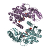 7cs4C 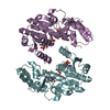 7cs5C  7cs6C 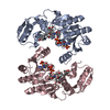 7cs7C 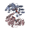 7cs8C  7cs9C  7csaC  7csbC  7cscC  7csdC  7cseC  7csfC  7csgC  7cshC  1qydS S: Starting model for refinement C: citing same article ( |
|---|---|
| Similar structure data |


-Links
 PDBj
PDBj- Assembly
| Deposited unit | 
| ||||||||||||
|---|---|---|---|---|---|---|---|---|---|---|---|---|---|
| 1 | 
| ||||||||||||
| 2 | 
| ||||||||||||
| 3 | 
| ||||||||||||
| Unit cell |
|
-Components
| #1: Protein | Mass: 35662.586 Da / Num. of mol.: 6 Source method: isolated from a genetically manipulated source Source: (gene. exp.) Isatis tinctoria (woad) / Production host:  #2: Water | ChemComp-HOH / |  Mass: 18.015 Da / Num. of mol.: 91 / Source method: isolated from a natural source / Formula: H2O Mass: 18.015 Da / Num. of mol.: 91 / Source method: isolated from a natural source / Formula: H2O |
|---|
-Experimental details
-Experiment
| Experiment | Method: X-RAY DIFFRACTION / Number of used crystals: 1 |
|---|
- Sample preparation
| Crystal | Density Matthews: 3.06 Å3/Da / Density % sol: 59.81 % |
|---|---|
| Crystal grow | Temperature: 293 K / Method: vapor diffusion, sitting drop / pH: 4 Details: 0.2M sodium citrate tribasic, 0.1 M sodium citrate/citric acid, pH 4.0, 20% PEG 3350 |
-Data collection
| Diffraction | Mean temperature: 100 K / Serial crystal experiment: N |
|---|---|
| Diffraction source | Source: SYNCHROTRON / Site: SSRF  / Beamline: BL17U1 / Wavelength: 0.9798 Å / Beamline: BL17U1 / Wavelength: 0.9798 Å |
| Detector | Type: ADSC QUANTUM 315r / Detector: CCD / Date: Dec 18, 2015 |
| Radiation | Protocol: SINGLE WAVELENGTH / Monochromatic (M) / Laue (L): M / Scattering type: x-ray |
| Radiation wavelength | Wavelength: 0.9798 Å / Relative weight: 1 |
| Reflection | Resolution: 2.688→31.399 Å / Num. obs: 70443 / % possible obs: 99.5 % / Redundancy: 6.7 % / Biso Wilson estimate: 66.1630245508 Å2 / Rmerge(I) obs: 0.093 / Net I/σ(I): 19.8 |
| Reflection shell | Resolution: 2.69→2.78 Å / Rmerge(I) obs: 0.764 / Num. unique obs: 6742 |
- Processing
| Software |
| |||||||||||||||||||||||||||||||||||||||||||||||||||||||||||||||||||||||||||||||||||||||||||||||||||||||||
|---|---|---|---|---|---|---|---|---|---|---|---|---|---|---|---|---|---|---|---|---|---|---|---|---|---|---|---|---|---|---|---|---|---|---|---|---|---|---|---|---|---|---|---|---|---|---|---|---|---|---|---|---|---|---|---|---|---|---|---|---|---|---|---|---|---|---|---|---|---|---|---|---|---|---|---|---|---|---|---|---|---|---|---|---|---|---|---|---|---|---|---|---|---|---|---|---|---|---|---|---|---|---|---|---|---|---|
| Refinement | Method to determine structure: MOLECULAR REPLACEMENT Starting model: 1QYD Resolution: 2.68800468428→31.3980458407 Å / SU ML: 0.348769881523 / Cross valid method: FREE R-VALUE / σ(F): 1.35349369364 / Phase error: 26.4830188047 Stereochemistry target values: GeoStd + Monomer Library + CDL v1.2
| |||||||||||||||||||||||||||||||||||||||||||||||||||||||||||||||||||||||||||||||||||||||||||||||||||||||||
| Solvent computation | Shrinkage radii: 0.9 Å / VDW probe radii: 1.11 Å / Solvent model: FLAT BULK SOLVENT MODEL | |||||||||||||||||||||||||||||||||||||||||||||||||||||||||||||||||||||||||||||||||||||||||||||||||||||||||
| Displacement parameters | Biso mean: 66.4566911034 Å2 | |||||||||||||||||||||||||||||||||||||||||||||||||||||||||||||||||||||||||||||||||||||||||||||||||||||||||
| Refinement step | Cycle: LAST / Resolution: 2.68800468428→31.3980458407 Å
| |||||||||||||||||||||||||||||||||||||||||||||||||||||||||||||||||||||||||||||||||||||||||||||||||||||||||
| Refine LS restraints |
| |||||||||||||||||||||||||||||||||||||||||||||||||||||||||||||||||||||||||||||||||||||||||||||||||||||||||
| LS refinement shell |
|
