 Movie
Movie Controller

[English] 日本語
 Yorodumi
- PDB-7col: Crystal structure of 5-ketofructose reductase complexed with NADPH -
+ Open data

- Basic information
| Entry | Database: PDB / ID: 7col | ||||||
|---|---|---|---|---|---|---|---|
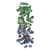
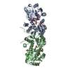
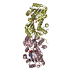
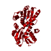
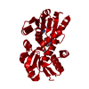
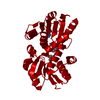
| Title | Crystal structure of 5-ketofructose reductase complexed with NADPH | ||||||
 Components Components | 5-ketofructose reductase | ||||||
 Keywords Keywords | OXIDOREDUCTASE / Gluconobacter / reductase / 5-ketofructose | ||||||
| Function / homology | Chem-NDP Function and homology information Function and homology information | ||||||
| Biological species |  Gluconobacter sp. (bacteria) Gluconobacter sp. (bacteria) | ||||||
| Method |  X-RAY DIFFRACTION / SYNCHROTRON / MOLECULAR REPLACEMENT / Resolution: 1.95 Å X-RAY DIFFRACTION / SYNCHROTRON / MOLECULAR REPLACEMENT / Resolution: 1.95 Å | ||||||
 Authors Authors | Hodoya, Y. / Noda, S. / Nguyen, T.M. / Kataoka, N. / Adachi, O. / Matsutani, M. / Matsushita, K. / Yakushi, T. / Goto, M. | ||||||
 Citation Citation | Journal: J.Bacteriol. / Year: 2021 Title: The 5-Ketofructose Reductase of Gluconobacter sp. Strain CHM43 Is a Novel Class in the Shikimate Dehydrogenase Family. Authors: Nguyen, T.M. / Goto, M. / Noda, S. / Matsutani, M. / Hodoya, Y. / Kataoka, N. / Adachi, O. / Matsushita, K. / Yakushi, T. | ||||||
| History |
|
- Structure visualization
| Structure viewer | Molecule: MolmilJmol/JSmol |
|---|
- Downloads & links
-Download
| PDBx/mmCIF format | 7col.cif.gz | 124.7 KB | Display | PDBx/mmCIF format |
|---|---|---|---|---|
| PDB format | pdb7col.ent.gz | 94.1 KB | Display | PDB format |
| PDBx/mmJSON format | 7col.json.gz | Tree view | PDBx/mmJSON format | |
| Others |  Other downloads Other downloads |
-Validation report
| Arichive directory | https://data.pdbj.org/pub/pdb/validation_reports/co/7colftp://data.pdbj.org/pub/pdb/validation_reports/co/7col | HTTPS FTP |
|---|
-Related structure data
| Related structure data |  7cokC  1nvtS S: Starting model for refinement C: citing same article ( |
|---|---|
| Similar structure data |
-Links
 PDBj
PDBj

- Assembly
| Deposited unit | 
| ||||||||
|---|---|---|---|---|---|---|---|---|---|
| 1 |
| ||||||||
| Unit cell |
|
-Components
| #1: Protein | Mass: 29514.707 Da / Num. of mol.: 2 Source method: isolated from a genetically manipulated source Source: (gene. exp.) Gluconobacter sp. (bacteria) / Strain: CHM43 / Plasmid: pBBR1MCS-4 / Production host: Gluconobacter sp. (bacteria) / Strain (production host): CHM43#2: Chemical |   Mass: 745.421 Da / Num. of mol.: 2 / Source method: obtained synthetically / Formula: C21H30N7O17P3 Mass: 745.421 Da / Num. of mol.: 2 / Source method: obtained synthetically / Formula: C21H30N7O17P3#3: Water | ChemComp-HOH / |  Mass: 18.015 Da / Num. of mol.: 399 / Source method: isolated from a natural source / Formula: H2O Mass: 18.015 Da / Num. of mol.: 399 / Source method: isolated from a natural source / Formula: H2OHas ligand of interest | N | |
|---|
-Experimental details
-Experiment
| Experiment | Method: X-RAY DIFFRACTION / Number of used crystals: 1 |
|---|
- Sample preparation
| Crystal | Density Matthews: 2.03 Å3/Da / Density % sol: 39.4 % |
|---|---|
| Crystal grow | Temperature: 295 K / Method: vapor diffusion, hanging drop / pH: 7.5 / Details: PEG 3350, HEPES-Na, Ammonium fluoride, MPD |
-Data collection
| Diffraction | Mean temperature: 95 K / Serial crystal experiment: N | |||||||||||||||||||||||||||||||||||||||||||||||||||||||||||||||||||||||||||||||||||||||||||||||||||||||||||||||||||||||||
|---|---|---|---|---|---|---|---|---|---|---|---|---|---|---|---|---|---|---|---|---|---|---|---|---|---|---|---|---|---|---|---|---|---|---|---|---|---|---|---|---|---|---|---|---|---|---|---|---|---|---|---|---|---|---|---|---|---|---|---|---|---|---|---|---|---|---|---|---|---|---|---|---|---|---|---|---|---|---|---|---|---|---|---|---|---|---|---|---|---|---|---|---|---|---|---|---|---|---|---|---|---|---|---|---|---|---|---|---|---|---|---|---|---|---|---|---|---|---|---|---|---|---|
| Diffraction source | Source: SYNCHROTRON / Site: Photon Factory  / Beamline: AR-NW12A / Wavelength: 1 Å / Beamline: AR-NW12A / Wavelength: 1 Å | |||||||||||||||||||||||||||||||||||||||||||||||||||||||||||||||||||||||||||||||||||||||||||||||||||||||||||||||||||||||||
| Detector | Type: ADSC QUANTUM 210r / Detector: CCD / Date: May 5, 2017 | |||||||||||||||||||||||||||||||||||||||||||||||||||||||||||||||||||||||||||||||||||||||||||||||||||||||||||||||||||||||||
| Radiation | Protocol: SINGLE WAVELENGTH / Monochromatic (M) / Laue (L): M / Scattering type: x-ray | |||||||||||||||||||||||||||||||||||||||||||||||||||||||||||||||||||||||||||||||||||||||||||||||||||||||||||||||||||||||||
| Radiation wavelength | Wavelength: 1 Å / Relative weight: 1 | |||||||||||||||||||||||||||||||||||||||||||||||||||||||||||||||||||||||||||||||||||||||||||||||||||||||||||||||||||||||||
| Reflection | Resolution: 1.95→96.169 Å / Num. all: 33451 / Num. obs: 33451 / % possible obs: 96.7 % / Redundancy: 3.2 % / Rpim(I) all: 0.039 / Rrim(I) all: 0.072 / Rsym value: 0.06 / Net I/av σ(I): 7.4 / Net I/σ(I): 13.1 / Num. measured all: 105580 | |||||||||||||||||||||||||||||||||||||||||||||||||||||||||||||||||||||||||||||||||||||||||||||||||||||||||||||||||||||||||
| Reflection shell | Diffraction-ID: 1
|
- Processing
| Software |
| |||||||||||||||||||||||||||||||||||||||||||||
|---|---|---|---|---|---|---|---|---|---|---|---|---|---|---|---|---|---|---|---|---|---|---|---|---|---|---|---|---|---|---|---|---|---|---|---|---|---|---|---|---|---|---|---|---|---|---|
| Refinement | Method to determine structure: MOLECULAR REPLACEMENT Starting model: 1NVT Resolution: 1.95→40.21 Å / Cor.coef. Fo:Fc: 0.939 / Cor.coef. Fo:Fc free: 0.906 / SU B: 5.273 / SU ML: 0.149 / SU R Cruickshank DPI: 0.2399 / Cross valid method: THROUGHOUT / σ(F): 0 / ESU R: 0.24 / ESU R Free: 0.2 / Stereochemistry target values: MAXIMUM LIKELIHOOD / Details: U VALUES : REFINED INDIVIDUALLY
| |||||||||||||||||||||||||||||||||||||||||||||
| Solvent computation | Ion probe radii: 0.8 Å / Shrinkage radii: 0.8 Å / VDW probe radii: 1.2 Å / Solvent model: MASK | |||||||||||||||||||||||||||||||||||||||||||||
| Displacement parameters | Biso max: 69.87 Å2 / Biso mean: 24.366 Å2 / Biso min: 12.32 Å2
| |||||||||||||||||||||||||||||||||||||||||||||
| Refinement step | Cycle: final / Resolution: 1.95→40.21 Å
| |||||||||||||||||||||||||||||||||||||||||||||
| Refine LS restraints |
| |||||||||||||||||||||||||||||||||||||||||||||
| LS refinement shell | Resolution: 1.95→2.001 Å / Rfactor Rfree error: 0 / Total num. of bins used: 20
|
