 Movie
Movie Controller

[English] 日本語
 Yorodumi
- PDB-3jyp: Quinate dehydrogenase from Corynebacterium glutamicum in complex ... -
+ Open data

- Basic information
| Entry | Database: PDB / ID: 3jyp | ||||||
|---|---|---|---|---|---|---|---|
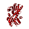
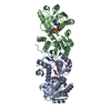
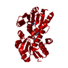
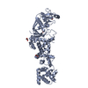
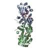
| Title | Quinate dehydrogenase from Corynebacterium glutamicum in complex with quinate and NADH | ||||||
 Components Components | Quinate/shikimate dehydrogenase | ||||||
 Keywords Keywords | OXIDOREDUCTASE / quinate dehyrogenase / ternary complex / quinate / NADH / Amino-acid biosynthesis / Aromatic amino acid biosynthesis / NAD | ||||||
| Function / homology |  Function and homology information Function and homology informationquinate/shikimate dehydrogenase (NAD+) / quinate 3-dehydrogenase (NAD+) activity / shikimate 3-dehydrogenase (NAD+) activity / shikimate 3-dehydrogenase (NADP+) activity / shikimate metabolic process / Oxidoreductases; Acting on the CH-OH group of donors; With NAD+ or NADP+ as acceptor / chorismate biosynthetic process / aromatic amino acid biosynthetic process / amino acid biosynthetic process / NAD+ binding ...quinate/shikimate dehydrogenase (NAD+) / quinate 3-dehydrogenase (NAD+) activity / shikimate 3-dehydrogenase (NAD+) activity / shikimate 3-dehydrogenase (NADP+) activity / shikimate metabolic process / Oxidoreductases; Acting on the CH-OH group of donors; With NAD+ or NADP+ as acceptor / chorismate biosynthetic process / aromatic amino acid biosynthetic process / amino acid biosynthetic process / NAD+ binding / NADP binding / cytosol Similarity search - Function | ||||||
| Biological species |  Corynebacterium glutamicum (bacteria) Corynebacterium glutamicum (bacteria) | ||||||
| Method |  X-RAY DIFFRACTION / SYNCHROTRON / MOLECULAR REPLACEMENT / Resolution: 1.16 Å X-RAY DIFFRACTION / SYNCHROTRON / MOLECULAR REPLACEMENT / Resolution: 1.16 Å | ||||||
 Authors Authors | Hoeppner, A. / Schomburg, D. / Niefind, K. | ||||||
 Citation Citation | Journal: Biol.Chem. / Year: 2013 Title: Enzyme-substrate complexes of the quinate/shikimate dehydrogenase from Corynebacterium glutamicum enable new insights in substrate and cofactor binding, specificity, and discrimination. Authors: Hoppner, A. / Schomburg, D. / Niefind, K. | ||||||
| History |
|
- Structure visualization
| Structure viewer | Molecule: MolmilJmol/JSmol |
|---|
- Downloads & links
-Download
| PDBx/mmCIF format | 3jyp.cif.gz | 136.7 KB | Display | PDBx/mmCIF format |
|---|---|---|---|---|
| PDB format | pdb3jyp.ent.gz | 106.3 KB | Display | PDB format |
| PDBx/mmJSON format | 3jyp.json.gz | Tree view | PDBx/mmJSON format | |
| Others |  Other downloads Other downloads |
-Validation report
| Arichive directory | https://data.pdbj.org/pub/pdb/validation_reports/jy/3jypftp://data.pdbj.org/pub/pdb/validation_reports/jy/3jyp | HTTPS FTP |
|---|
-Related structure data
| Related structure data |  3jyoSC  3jyqC S: Starting model for refinement C: citing same article ( |
|---|---|
| Similar structure data |


-Links
 PDBj
PDBj- Assembly
| Deposited unit | 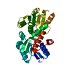
| ||||||||
|---|---|---|---|---|---|---|---|---|---|
| 1 | 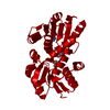
| ||||||||
| Unit cell |
|
-Components
| #1: Protein | Mass: 29724.580 Da / Num. of mol.: 1 / Fragment: QDH Source method: isolated from a genetically manipulated source Source: (gene. exp.) Corynebacterium glutamicum (bacteria) / Strain: ATCC13032 / Gene: aroE, cg0504, Cgl0424 / Plasmid: pNHis / Production host: References: UniProt: Q9X5C9, quinate/shikimate dehydrogenase (NAD+), Oxidoreductases; Acting on the CH-OH group of donors; With NAD+ or NADP+ as acceptor |
|---|---|
| #2: Chemical | ChemComp-NAD /   Mass: 663.425 Da / Num. of mol.: 1 / Source method: obtained synthetically / Formula: C21H27N7O14P2 / Comment: NAD*YM Mass: 663.425 Da / Num. of mol.: 1 / Source method: obtained synthetically / Formula: C21H27N7O14P2 / Comment: NAD*YM |
| #3: Chemical | ChemComp-QIC / (  Mass: 192.167 Da / Num. of mol.: 1 / Source method: obtained synthetically / Formula: C7H12O6 Mass: 192.167 Da / Num. of mol.: 1 / Source method: obtained synthetically / Formula: C7H12O6 |
| #4: Water | ChemComp-HOH /  Mass: 18.015 Da / Num. of mol.: 369 / Source method: isolated from a natural source / Formula: H2O Mass: 18.015 Da / Num. of mol.: 369 / Source method: isolated from a natural source / Formula: H2O |
-Experimental details
-Experiment
| Experiment | Method: X-RAY DIFFRACTION / Number of used crystals: 1 |
|---|
- Sample preparation
| Crystal | Density Matthews: 2.29 Å3/Da / Density % sol: 46.28 % |
|---|---|
| Crystal grow | Temperature: 293 K / Method: vapor diffusion, sitting drop / pH: 9.5 Details: 24 % PEG 6000, 400 mM calcium chloride, 100 mM Tris/HCl, pH 9.5, VAPOR DIFFUSION, SITTING DROP, temperature 293K |
-Data collection
| Diffraction | Mean temperature: 100 K |
|---|---|
| Diffraction source | Source: SYNCHROTRON / Site: EMBL/DESY, HAMBURG  / Beamline: X12 / Wavelength: 0.9 Å / Beamline: X12 / Wavelength: 0.9 Å |
| Detector | Type: MAR scanner 345 mm plate / Detector: IMAGE PLATE / Date: Oct 5, 2007 |
| Radiation | Protocol: SINGLE WAVELENGTH / Monochromatic (M) / Laue (L): M / Scattering type: x-ray |
| Radiation wavelength | Wavelength: 0.9 Å / Relative weight: 1 |
| Reflection | Resolution: 1.16→30 Å / Num. all: 92402 / Num. obs: 88244 / % possible obs: 95.9 % / Observed criterion σ(F): 0 / Observed criterion σ(I): 0 / Redundancy: 6.3 % / Biso Wilson estimate: 17.48 Å2 / Rmerge(I) obs: 0.081 / Rsym value: 0.081 / Net I/σ(I): 26.87 |
| Reflection shell | Resolution: 1.16→1.2 Å / Redundancy: 4.3 % / Rmerge(I) obs: 0.4 / Mean I/σ(I) obs: 4.19 / Num. unique all: 8491 / Rsym value: 0.4 / % possible all: 92.5 |
- Processing
| Software |
| |||||||||||||||||||||||||||||||||||||||||||||||||||||||
|---|---|---|---|---|---|---|---|---|---|---|---|---|---|---|---|---|---|---|---|---|---|---|---|---|---|---|---|---|---|---|---|---|---|---|---|---|---|---|---|---|---|---|---|---|---|---|---|---|---|---|---|---|---|---|---|---|
| Refinement | Method to determine structure: MOLECULAR REPLACEMENT Starting model: 3jyo Resolution: 1.16→30 Å / Cross valid method: THROUGHOUT / σ(F): 0 / σ(I): 0
| |||||||||||||||||||||||||||||||||||||||||||||||||||||||
| Displacement parameters | Biso mean: 17.48 Å2 | |||||||||||||||||||||||||||||||||||||||||||||||||||||||
| Refinement step | Cycle: LAST / Resolution: 1.16→30 Å
| |||||||||||||||||||||||||||||||||||||||||||||||||||||||
| LS refinement shell |
|
