 Movie
Movie Controller

[English] 日本語
 Yorodumi
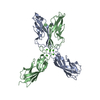
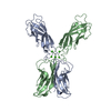
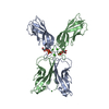
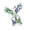
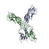
- PDB-7cme: Crystal structure of human P-cadherin MEC12 (X dimer) in complex ... -
+ Open data

- Basic information
| Entry | Database: PDB / ID: 7cme | ||||||
|---|---|---|---|---|---|---|---|
| Title | Crystal structure of human P-cadherin MEC12 (X dimer) in complex with 2-(5-chloro-2-methyl-1H-indol-3-yl)ethan-1-amine (inhibitor) | ||||||
 Components Components | Cadherin-3 | ||||||
 Keywords Keywords | CELL ADHESION / inhibitor / complex | ||||||
| Function / homology |  Function and homology information Function and homology information: / negative regulation of timing of catagen / positive regulation of melanosome transport / hair cycle process / positive regulation of keratinocyte proliferation / positive regulation of melanin biosynthetic process / calcium-dependent cell-cell adhesion / cell-cell adhesion mediated by cadherin / adherens junction organization / catenin complex ...: / negative regulation of timing of catagen / positive regulation of melanosome transport / hair cycle process / positive regulation of keratinocyte proliferation / positive regulation of melanin biosynthetic process / calcium-dependent cell-cell adhesion / cell-cell adhesion mediated by cadherin / adherens junction organization / catenin complex / cell-cell junction assembly / retina homeostasis / Adherens junctions interactions / positive regulation of insulin-like growth factor receptor signaling pathway / keratinization / homophilic cell-cell adhesion / visual perception / adherens junction / negative regulation of transforming growth factor beta receptor signaling pathway / beta-catenin binding / cell morphogenesis / cell junction / cell migration / positive regulation of canonical Wnt signaling pathway / cell adhesion / cadherin binding / calcium ion binding / positive regulation of gene expression / plasma membrane / cytoplasm Similarity search - Function | ||||||
| Biological species |  Homo sapiens (human) Homo sapiens (human) | ||||||
| Method |  X-RAY DIFFRACTION / SYNCHROTRON / MOLECULAR REPLACEMENT / molecular replacement / Resolution: 2.45 Å X-RAY DIFFRACTION / SYNCHROTRON / MOLECULAR REPLACEMENT / molecular replacement / Resolution: 2.45 Å | ||||||
 Authors Authors | Senoo, A. / Ito, S. / Ueno, G. / Nagatoishi, S. / Tsumoto, K. | ||||||
 Citation Citation | Journal: Commun Biol / Year: 2021 Title: Regulation of cadherin dimerization by chemical fragments as a trigger to inhibit cell adhesion Authors: Senoo, A. / Ito, S. / Nagatoishi, S. / Saito, Y. / Ueno, G. / Kuroda, D. / Yoshida, K. / Tashima, T. / Kudo, S. / Sando, S. / Tsumoto, K. | ||||||
| History |
|
- Structure visualization
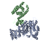
| Structure viewer | Molecule: MolmilJmol/JSmol |
|---|
- Downloads & links
-Download
| PDBx/mmCIF format | 7cme.cif.gz | 101.8 KB | Display | PDBx/mmCIF format |
|---|---|---|---|---|
| PDB format | pdb7cme.ent.gz | 76 KB | Display | PDB format |
| PDBx/mmJSON format | 7cme.json.gz | Tree view | PDBx/mmJSON format | |
| Others |  Other downloads Other downloads |
-Validation report
| Arichive directory | https://data.pdbj.org/pub/pdb/validation_reports/cm/7cmeftp://data.pdbj.org/pub/pdb/validation_reports/cm/7cme | HTTPS FTP |
|---|
-Related structure data
| Related structure data |  7cmfC  4zmqS S: Starting model for refinement C: citing same article ( |
|---|---|
| Similar structure data |

-Links
 PDBj
PDBj

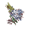
- Assembly
| Deposited unit | 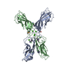
| ||||||||
|---|---|---|---|---|---|---|---|---|---|
| 1 |
| ||||||||
| Unit cell |
|
-Components
| #1: Protein | Mass: 23422.012 Da / Num. of mol.: 2 Source method: isolated from a genetically manipulated source Source: (gene. exp.) Homo sapiens (human) / Gene: CDH3, CDHP / Production host:  #2: Chemical |   Mass: 78.133 Da / Num. of mol.: 2 / Source method: obtained synthetically / Formula: C2H6OS / Comment: DMSO, precipitant*YM Mass: 78.133 Da / Num. of mol.: 2 / Source method: obtained synthetically / Formula: C2H6OS / Comment: DMSO, precipitant*YM#3: Chemical |   Mass: 208.687 Da / Num. of mol.: 3 / Source method: obtained synthetically / Formula: C11H13ClN2 / Feature type: SUBJECT OF INVESTIGATION Mass: 208.687 Da / Num. of mol.: 3 / Source method: obtained synthetically / Formula: C11H13ClN2 / Feature type: SUBJECT OF INVESTIGATION#4: Chemical | ChemComp-CA /   Mass: 40.078 Da / Num. of mol.: 6 / Source method: obtained synthetically / Formula: Ca Mass: 40.078 Da / Num. of mol.: 6 / Source method: obtained synthetically / Formula: Ca#5: Water | ChemComp-HOH / |  Mass: 18.015 Da / Num. of mol.: 29 / Source method: isolated from a natural source / Formula: H2O Mass: 18.015 Da / Num. of mol.: 29 / Source method: isolated from a natural source / Formula: H2OHas ligand of interest | Y | |
|---|
-Experimental details
-Experiment
| Experiment | Method: X-RAY DIFFRACTION / Number of used crystals: 1 |
|---|
- Sample preparation
| Crystal | Density Matthews: 4.56 Å3/Da / Density % sol: 73.01 % |
|---|---|
| Crystal grow | Temperature: 293 K / Method: vapor diffusion, sitting drop Details: 0.17M Sodium acetate trihydrate, 0.085M TRIS pH 8.5, w/v 25.5% PEG 4000, 15% v/v Glycerol |
-Data collection
| Diffraction | Mean temperature: 100 K / Serial crystal experiment: N | ||||||||||||||||||||||||||||||||||||||||||||||||||||||||||||||||||||||||||||||||||||||||||||||||||||
|---|---|---|---|---|---|---|---|---|---|---|---|---|---|---|---|---|---|---|---|---|---|---|---|---|---|---|---|---|---|---|---|---|---|---|---|---|---|---|---|---|---|---|---|---|---|---|---|---|---|---|---|---|---|---|---|---|---|---|---|---|---|---|---|---|---|---|---|---|---|---|---|---|---|---|---|---|---|---|---|---|---|---|---|---|---|---|---|---|---|---|---|---|---|---|---|---|---|---|---|---|---|
| Diffraction source | Source: SYNCHROTRON / Site: SPring-8  / Beamline: BL26B2 / Wavelength: 1 Å / Beamline: BL26B2 / Wavelength: 1 Å | ||||||||||||||||||||||||||||||||||||||||||||||||||||||||||||||||||||||||||||||||||||||||||||||||||||
| Detector | Type: RAYONIX MX225-HS / Detector: CCD / Date: Jul 24, 2019 | ||||||||||||||||||||||||||||||||||||||||||||||||||||||||||||||||||||||||||||||||||||||||||||||||||||
| Radiation | Protocol: SINGLE WAVELENGTH / Monochromatic (M) / Laue (L): M / Scattering type: x-ray | ||||||||||||||||||||||||||||||||||||||||||||||||||||||||||||||||||||||||||||||||||||||||||||||||||||
| Radiation wavelength | Wavelength: 1 Å / Relative weight: 1 | ||||||||||||||||||||||||||||||||||||||||||||||||||||||||||||||||||||||||||||||||||||||||||||||||||||
| Reflection | Resolution: 2.45→45.04 Å / Num. obs: 31971 / % possible obs: 99.9 % / Redundancy: 7.38 % / Biso Wilson estimate: 68.736 Å2 / CC1/2: 0.999 / Rmerge(I) obs: 0.071 / Rrim(I) all: 0.076 / Χ2: 0.862 / Net I/σ(I): 19.21 / Num. measured all: 235947 | ||||||||||||||||||||||||||||||||||||||||||||||||||||||||||||||||||||||||||||||||||||||||||||||||||||
| Reflection shell | Diffraction-ID: 1
|
-Phasing
| Phasing | Method: molecular replacement | ||||||
|---|---|---|---|---|---|---|---|
| Phasing MR |
|
- Processing
| Software |
| ||||||||||||||||||||||||||||||||||||||||||||||||||||||||||||||||||||||||||||||
|---|---|---|---|---|---|---|---|---|---|---|---|---|---|---|---|---|---|---|---|---|---|---|---|---|---|---|---|---|---|---|---|---|---|---|---|---|---|---|---|---|---|---|---|---|---|---|---|---|---|---|---|---|---|---|---|---|---|---|---|---|---|---|---|---|---|---|---|---|---|---|---|---|---|---|---|---|---|---|---|
| Refinement | Method to determine structure: MOLECULAR REPLACEMENT Starting model: 4zmq Resolution: 2.45→45.04 Å / SU ML: 0.4 / Cross valid method: THROUGHOUT / σ(F): 1.33 / Phase error: 28.42 / Stereochemistry target values: ML
| ||||||||||||||||||||||||||||||||||||||||||||||||||||||||||||||||||||||||||||||
| Solvent computation | Shrinkage radii: 0.9 Å / VDW probe radii: 1.11 Å / Solvent model: FLAT BULK SOLVENT MODEL | ||||||||||||||||||||||||||||||||||||||||||||||||||||||||||||||||||||||||||||||
| Displacement parameters | Biso max: 166.93 Å2 / Biso mean: 76.5952 Å2 / Biso min: 38.39 Å2 | ||||||||||||||||||||||||||||||||||||||||||||||||||||||||||||||||||||||||||||||
| Refinement step | Cycle: final / Resolution: 2.45→45.04 Å
| ||||||||||||||||||||||||||||||||||||||||||||||||||||||||||||||||||||||||||||||
| LS refinement shell | Refine-ID: X-RAY DIFFRACTION / Rfactor Rfree error: 0 / Total num. of bins used: 12 / % reflection obs: 100 %
|
